. 2016 Sep;30(9):3155-70.
doi: 10.1096/fj.201600257R. Epub 2016 Jun 17.

Cold/menthol TRPM8 receptors initiate the cold-shock response and protect germ cells from cold-shock-induced oxidation

Affiliations

Cold/menthol TRPM8 receptors initiate the cold-shock response and protect germ cells from cold-shock-induced oxidation

Anne-Sophie Borowiec et al. FASEB J. 2016 Sep.

Abstract

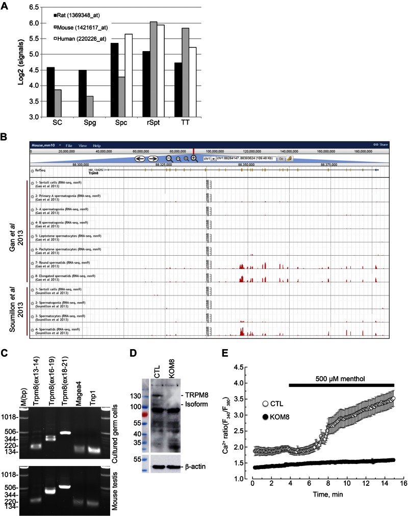
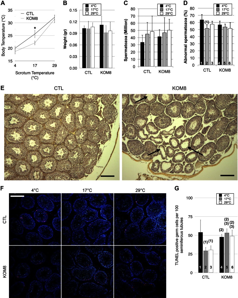
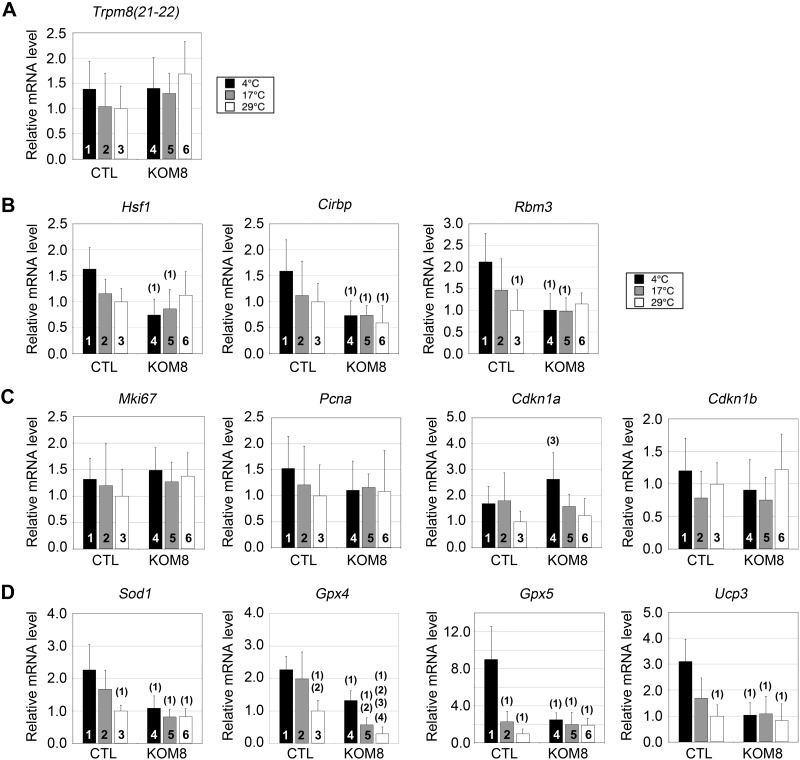
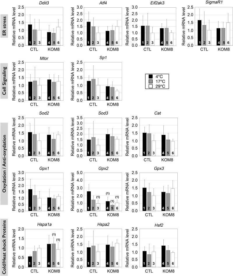
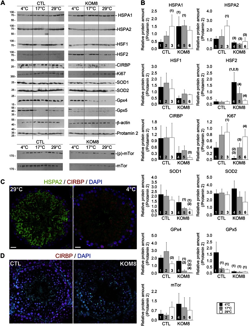
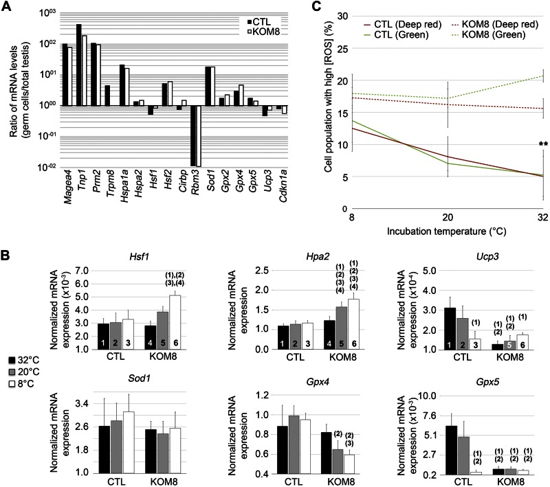
Testes of most male mammals present the particularity of being externalized from the body and are consequently slightly cooler than core body temperature (4-8°C below). Although, hypothermia of the testis is known to increase germ cells apoptosis, little is known about the underlying molecular mechanisms, including cold sensors, transduction pathways, and apoptosis triggers. In this study, using a functional knockout mouse model of the cold and menthol receptors, dubbed transient receptor potential melastatine 8 (TRPM8) channels, we found that TRPM8 initiated the cold-shock response by differentially modulating cold- and heat-shock proteins. Besides, apoptosis of germ cells increased in proportion to the cooling level in control mice but was independent of temperature in knockout mice. We also observed that the rate of germ cell death correlated positively with the reactive oxygen species level and negatively with the expression of the detoxifying enzymes. This result suggests that the TRPM8 sensor is a key determinant of germ cell fate under hypothermic stimulation.-Borowiec, A.-S., Sion, B., Chalmel, F., Rolland, A. D., Lemonnier, L., De Clerck, T., Bokhobza, A., Derouiche, S., Dewailly, E., Slomianny, C., Mauduit, C., Benahmed, M., Roudbaraki, M., Jégou, B., Prevarskaya, N., Bidaux, G. Cold/menthol TRPM8 receptors initiate the cold-shock response and protect germ cells from cold-shock-induced oxidation.

Keywords: apoptosis; hypothermia; spermatogenesis.


Figures

Figure 1.
Figure 1.
TRPM8 channels are expressed and functional in mouse germ cells. A) Normalized signal intensities (y axis, log2-transformed) of Trpm8 are shown in the different testicular cell types (x axis) in Sertoli cells (SC), spermatogonia (Spg), spermatocytes (Spc), round spermatids (rSpt), and total testis (TT) of 3 mammalian species: rat, mouse, and human. B) Gene structure is shown for Trpm8 and histograms of the numbers of RNA-seq reads that aligned the corresponding genomic locations across the different samples from Gan et al. (21) and Soumillon et al. (22) (y-axis ranges from 0 to 40) (adapted from The ReproGenomics Viewer; http://rgv.genouest.org/publication.html). C) PCR detection of different regions of Trpm8 in cultured germ cells (top) and in whole extracts of mouse testis (bottom). PCR fragments were amplified from exon X to Y and reported as Trpm8(exX-Y). Melanoma antigen family A-4 (Magea 4) and transition protein 1 (Tnp1) were used as reporters of spermatogonia and spermatids respectively. D) Immunoblot analysis reveals detection of full-length TRPM8 (130 kDa) and a 105 kDa TRPM8 isoform, as well, in total protein extract of CTL mouse testis, but not in TRPM8-KO (KOM8) mouse testis. E) Calcium imaging experiments realized with Fura2-AM fluorescent probe show an increased cytosolic Ca2+ concentration in 2 d isolated Trpm8+/+ mouse germ cells (CTL; n = 20) after addition of 500 µM menthol. No Ca2+ variation was detected in germ cells of Trpm8−/− mouse line (KOM8; n = 83).
Figure 2.
Figure 2.
TRPM8 channel expression protects germ cells from apoptosis at rest and during mild, but not noxious, cold exposure. A) Trpm8+/+ (CTL) and Trpm8−/− (KOM8) mice were subjected to a 45 min cold shock, and both body and scrotum temperatures were measured concomitantly. Mild cold (17°C) revealed an impaired thermogenesis of KOM8 testis, whereas no alteration appeared under noxious cold conditions (4°C). BD) Testicular weight (B), spermatozoa concentration (C), and percentage of abnormal spermatozoa (D) in CTL and KOM8 mouse testes subjected to cold shocks of different magnitudes did not show any significant difference 3 d after treatment. Statistical significance was assumed when P < 0.05 and is shown above the tested column as the number of the column it is paired to. E) Anatomopathological analysis revealed the presence of near-empty seminiferous tubules (arrows) in trichromatic-stained paraffin-embedded sections of KOM8 testis of nonstimulated mice. Scale bars, 200 µm. F) TUNEL with nucleotides coupled to tetramethyl rhodamine (red) revealed apoptotic germ cells. Nuclei were counterstained with DAPI (blue). Apoptotic cells were counted and normalized by 100 seminiferous tubules. Scale bar, 200 µm. G) Histogram shows statistical analysis of the counting of apoptotic germ cells. Values are means ± sd for CTL (n = 6) and KOM8 (n = 6) mice. Statistical analysis was performed by 1-way ANOVA.
Figure 3.
Figure 3.
Screening of specific gene networks reveals the TRPM8-dependence of genes encoding cold-shock proteins and antioxidant enzymes. A) Real-time PCR showed no significant variation in Trpm8 gene expression (exons 21–22) in CTL and KOM8 mice subjected to cold shocks of different amplitudes (scrotum temperatures were 29, 17, and 4°C). BD) mRNA quantification was also performed for the following heat- and cold-shock factors (B): heat-shock factor 1 (Hsf1), cold-inducible RNA binding protein (Cirbp) and RNA binding motif (RNP1, RRM) protein 3 (Rbm3); for proliferation markers (C): antigen identified by monoclonal antibody Ki 67 (Mki67) and proliferating cell nuclear antigen (Pcna); inhibitors of cell cycle: cyclin-dependent kinase inhibitor 1A and 1B (Cdkn1a and Cdkn1b, respectively) also known as p21cip1/waf1 and p27kip; and for antioxidant enzymes (D): SOD1, Cu2+/Zn2+ (Sod1, Gpx4, and Gpx5, respectively) and UCP3 (Ucp3). Values are presented as means ± sd for CTL (n = 5) and KOM8 (n = 5) mice. Statistical significance confirmed with 1-way ANOVA when P < 0.05 and is indicated above the tested column as the number of the CTL column it is paired to.
Figure 4.
Figure 4.
Real-time PCR screening of several genes families in mouse testis 3 d after cold shocks. As in Fig. 3, graphs show real-time PCR quantification of gene expression of ER stress marker: DNA-damage-inducible transcript 3 (Ddit3), activating transcription factor 4 (Atf4), eukaryotic translation initiation factor 2-α kinase 3 (Eif2ak3), σ nonopioid intracellular receptor 1 (Sigmar1); cell signaling markers: mechanistic target of rapamycin (serine/threonine kinase) (Mtor) and Sp1 transcription factor (Sp1); oxidation/anti-oxidation enzymes: Sod2 and -3, respectively, catalase, Gpx1, -2, and -3 respectively; cold/heat-shock induced genes: heat-shock protein -1A and 2A (Hspa1a and -2, respectively) and heat shock factor 2 (Hsf2). Statistical significance at P < 0.05 was confirmed with 1-way ANOVA and is shown above the tested column as the number of the CTL column it is paired to.
Figure 5.
Figure 5.
TRPM8 channels participate in the regulation of specific proteins expression in a cold-dependent and cold-independent way. A) Immunoblot analysis showing the variation of key protein expression in testis of 4 CTL and 4 KOM8 mice 3 d after cold shock (scrotum temperatures were 29, 17, or 4°C). β-Actin and Prm2 were used as invariant reporters. FKBP-rapamycin–associated protein was detected in its nonphosphorylated and phosphorylated (Ser2448) forms. B) Quantification of the protein levels and statistical significance are reported in (B). Protein amount was normalized with Prm2, which appeared much more stable than actin or GAPDH (not shown). Values are presented as means ± sd for CTL (n = 4) and KOM8 (n = 4) mice. Statistical significance was assumed when P < 0.05 and is shown above the tested column as the number of the CTL column it is paired to. C, D) Immunohistofluorescence confirmed the decreased expression of HSPA2 (C, green) in CTL germ cells subjected to hypothermia (C, right) compared to CTL (left). CIRBP (red) expression appears stable in both conditions. However, CIRBP was down-regulated in KOM8 testis (D, right) when compared to CTL (D, left). Nuclei were counterstained with DAPI. Scale bars, 20 µm. Statistical analysis was achieved with 1-way ANOVA.
Figure 6.
Figure 6.
Cold-induced TRPM8 channel activity triggers cold shock response and ROS production. A) Expression level of genes of interest in freshly isolated germ cells from either Trpm8+/+ (CTL; black bars) or Trpm8−/− mouse line (KOM8; white bars). Values are expressed as the ratio of gene expression on total mouse testis expression. Rbm3 was the sole gene with drastically decreased expression in isolated germ cells. Conversely, Hspa1a, Hsf2, Sod1, and Gpx2 exhibited a preferential expression in germ cells. B) Real-time based quantification of mRNA levels of Hsf1, Hspa2, Ucp3, Sod1, Gpx4, and Gpx5 in freshly isolated CTL or KOM8 mouse germ cells subjected to a 1-h incubation at 8, 20, or 32°C. Statistical significance (1-way ANOVA) was assumed when P < 0.05 and is shown above the tested column as the number of the CTL column it is paired to. C) Freshly isolated mouse germ cells were loaded with the nuclear CellROX Green Reagent and the cytosolic/mitochondrial CellROX deep Red Reagent before being subjected to a 1 h cold shock (8°C and 20°C) or kept at CTL temperature (32°C). Cytometer analysis reveals a significant cold-dependent accumulation of ROS in CTL germ cells, whereas stable ROS concentration was observed in KOM8 germ cells. Values are expressed as means ± sd for CTL (n = 4) and KOM8 (n = 4) mice. Statistical analysis was achieved with a t test.

References

    1. Chowdhury A. K., Steinberger E. (1970) Early changes in the germinal epithelium of rat testes following exposure to heat. J. Reprod. Fertil. 22, 205–212 - PubMed
    1. Macdonald J., Harrison R. G. (1954) Effect of low temperatures on rat spermatogenesis. Fertil. Steril. 5, 205–216 - PubMed
    1. Blanco-Rodríguez J., Martínez-García C. (1997) Mild hypothermia induces apoptosis in rat testis at specific stages of the seminiferous epithelium. J. Androl. 18, 535–539 - PubMed
    1. Zhang Z., Short R. V., Meehan T., De Kretser D. M., Renfree M. B., Loveland K. L. (2004) Functional analysis of the cooled rat testis. J. Androl. 25, 57–68 - PubMed
    1. Yazawa T., Nakayama Y., Fujimoto K., Matsuda Y., Abe K., Kitano T., Abé S., Yamamoto T. (2003) Abnormal spermatogenesis at low temperatures in the Japanese red-bellied newt, Cynops pyrrhogaster: possible biological significance of the cessation of spermatocytogenesis. Mol. Reprod. Dev. 66, 60–66 - PubMed

Publication types

LinkOut - more resources