. 2016 Aug 1;143(15):2716-23.
doi: 10.1242/dev.123935. Epub 2016 Jun 17.

Functional analysis of AEBP2, a PRC2 Polycomb protein, reveals a Trithorax phenotype in embryonic development and in ESCs

Affiliations

Functional analysis of AEBP2, a PRC2 Polycomb protein, reveals a Trithorax phenotype in embryonic development and in ESCs

Anne Grijzenhout et al. Development. .

Abstract

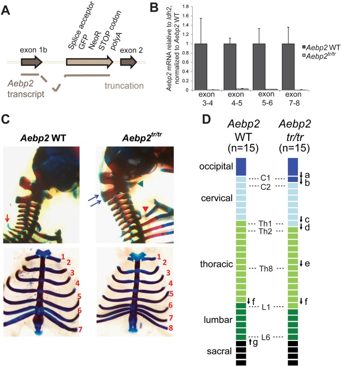
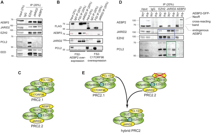
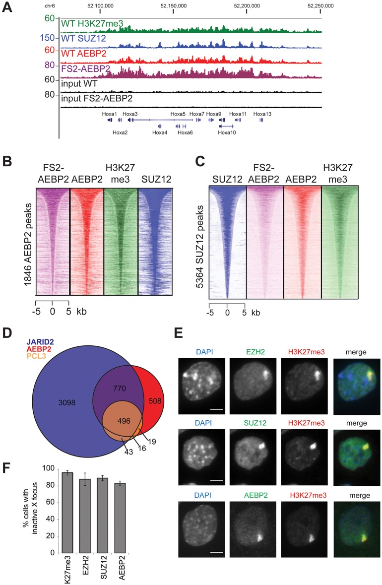
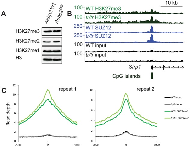
The Polycomb repressive complexes PRC1 and PRC2 are key mediators of heritable gene silencing in multicellular organisms. Here, we characterise AEBP2, a known PRC2 co-factor which, in vitro, has been shown to stimulate PRC2 activity. We show that AEBP2 localises specifically to PRC2 target loci, including the inactive X chromosome. Proteomic analysis confirms that AEBP2 associates exclusively with PRC2 complexes. However, analysis of embryos homozygous for a targeted mutation of Aebp2 unexpectedly revealed a Trithorax phenotype, normally linked to antagonism of Polycomb function. Consistent with this, we observe elevated levels of PRC2-mediated histone H3K27 methylation at target loci in Aebp2 mutant embryonic stem cells (ESCs). We further demonstrate that mutant ESCs assemble atypical hybrid PRC2 subcomplexes, potentially accounting for enhancement of Polycomb activity, and suggesting that AEBP2 normally plays a role in defining the mutually exclusive composition of PRC2 subcomplexes.

Keywords: AEBP2; Chromatin; Mouse; PRC2; Polycomb; Trithorax.


Conflict of interest statement

The authors declare no competing or financial interests.

Figures

Fig. 1.
Fig. 1.
Aebp2 truncation leads to perinatal lethality and anterior homeotic transformations. (A) Insertion of the splice acceptor cassette in front of exon 2 leads to trapping of the transcript and a protein product that contains the first 217 amino acids (aa) of AEBP2, encoded in exon 1b, fused to green fluorescent protein (GFP) and aminoglycoside 3′ phosphotransferase (NeoR). (B) The levels of Aebp2 mRNA transcripts containing exons downstream of the trapping cassette are severely reduced in Aebp2tr/tr mESCs compared with the parental WT Aebp2 mESCs. Error bars indicate s.d. (C) Lateral views of the occipito-cervical region (top panels) and ventral views of the rib cage (bottom) of Aebp2 WT (left) and Aebp2tr/tr (right) foetuses. In Aebp2tr/tr foetuses, the ventral region of the atlas (C1, indicated by a green arrowhead) is fused to that of the axis (C2). Ventral ossification centre of the atlas was laterally expanded and acquired similar features to occipital bone. The dorsal region of the axis was cranio-caudally expanded and partially bifurcated (indicated by blue arrows). The proximal region of the rib associated with the first thoracic vertebra (Th1) was not formed (indicated by a red arrowhead). The prominent dorsal process, which is associated with the Th2 in the Aebp2 WT (indicated by a red arrow), is not formed in the Aebp2tr/tr. Bottom panel also shows association of the 8th rib to the sternum. (D) Schematic summary for homeotic transformations of the axis in the Aebp2tr/tr foetuses.
Fig. 2.
Fig. 2.
AEBP2 is part of the PRC2.2 subcomplex and defines accessory substoichiometric subunit association. (A) Immunoprecipitation of endogenous PRC2 subunits EZH2, JARID2 and AEBP2 in E14 mESCs. Bands of different molecular weight seen in the immunoblots of PCL2 and EED represent different isoforms of the proteins. (B) Immunoblot of co-immunoprecipitation of FS2-tagged AEBP2 (PRC2.2) and FS2-tagged C17ORF96 (PRC2.1) using either the Flag-2xStrepII (FS2) antibody, or the JARID2 antibody, illustrating that each complex associates with different PRC2 factors. The red asterisks indicate cross-reacting bands (see also Fig. S2B). (C) Schematic of PRC2.1 and PRC2.2 subcomplexes. (D) Immunoblot analysis of co-immunoprecipitation experiments to analyse the composition of PRC2 complexes. The blue box highlights the lanes that show a decreased association of EZH2 and JARID2 in Aebp2tr/tr mESCs, and the green box highlights the lanes showing an increased association of PCL2 and JARID2 upon loss of AEBP2. (E) Model of altered complex composition after AEBP2 depletion.
Fig. 3.
Fig. 3.
AEBP2 is enriched at PRC2 target sites. (A) ChIP-seq profile for SUZ12, H3K27me3, AEBP2 and input, and FS2-AEBP2 and input at the HoxA locus. Two repeats of ChIP-seq for the endogenous proteins were performed. Traces from one repeat are shown. (B) Heat-map analysis of AEBP2 peaks (1846), showing ChIP-seq signal for FS2-tagged AEBP2, AEBP2, H3K27me3 and SUZ12 at a 10 kb region centred over the AEBP2 peaks. (C) Heat-map analysis of SUZ12 peaks (5364), showing ChIP-seq signal for FS2-tagged AEBP2, AEBP2 and SUZ12 at a 10 kb region centred over the SUZ12 peaks. AEBP2 and FS2-AEBP2 are enriched at SUZ12 sites. (D) Overlap of peak datasets of JARID2 (Peng et al., 2009), AEBP2 and PCL3 (Brien et al., 2012). (E) Immunofluorescence images indicating overlap of H3K27me3, which marks the inactive X chromosome in trophoblast stem cells, and the PRC2 proteins EZH2, SUZ12 and AEBP2. Scale bar: 5 µm. (F) Quantification of the number of trophoblast stem cells observed with inactive X focus. A minimum of 300 cells were counted in three biological repeats. Error bars indicate s.d.
Fig. 4.
Fig. 4.
Aebp2tr/tr cells show a small increase in H3K27me3 at PRC2 target sites. (A) Immunoblot of global H3K27me0/1/2/3 levels in Aebp2 WT and Aebp2tr/tr mESCs. (B) ChIP-seq profile for H3K27me3, SUZ12 and input in Aebp2 WT and Aebp2tr/tr mESCs at the Sfrp1 gene. (C) A metaplot of H3K27me3 ChIP-seq signal at AEBP2 sites in Aebp2 WT and Aebp2tr/tr mESCs plotted across a 10 kb window for two repeats of the ChIP-seq experiment.

References

    1. Akasaka T., Kanno M., Balling R., Mieza M. A., Taniguchi M. and Koseki H. (1996). A role for mel-18, a Polycomb group-related vertebrate gene, during the anteroposterior specification of the axial skeleton. Development 122, 1513-1522. - PubMed
    1. Alekseyenko A. A., Gorchakov A. A., Kharchenko P. V. and Kuroda M. I. (2014). Reciprocal interactions of human C10orf12 and C17orf96 with PRC2 revealed by BioTAP-XL cross-linking and affinity purification. Proc. Natl. Acad. Sci. USA 111, 2488-2493. 10.1073/pnas.1400648111 - DOI - PMC - PubMed
    1. Ballaré C., Lange M., Lapinaite A., Martin G. M., Morey L., Pascual G., Liefke R., Simon B., Shi Y., Gozani O. et al. (2012). Phf19 links methylated Lys36 of histone H3 to regulation of Polycomb activity. Nat. Struct. Mol. Biol. 19, 1257-1265. 10.1038/nsmb.2434 - DOI - PMC - PubMed
    1. Blackledge N. P., Farcas A. M., Kondo T., King H. W., McGouran J. F., Hanssen L. L. P., Ito S., Cooper S., Kondo K., Koseki Y. et al. (2014). Variant PRC1 complex-dependent H2A ubiquitylation drives PRC2 recruitment and polycomb domain formation. Cell 157, 1445-1459. 10.1016/j.cell.2014.05.004 - DOI - PMC - PubMed
    1. Brien G. L., Gambero G., O'Connell D. J., Jerman E., Turner S. A., Egan C. M., Dunne E. J., Jurgens M. C., Wynne K., Piao L. et al. (2012). Polycomb PHF19 binds H3K36me3 and recruits PRC2 and demethylase NO66 to embryonic stem cell genes during differentiation. Nat. Struct. Mol. Biol. 19, 1273-1281. 10.1038/nsmb.2449 - DOI - PubMed