. 2016 Aug;11(2):129-34.
doi: 10.1007/s11751-016-0257-3. Epub 2016 Jun 18.

Percutaneous rotational osteotomy of the femur utilizing an intramedullary rod

Affiliations

Percutaneous rotational osteotomy of the femur utilizing an intramedullary rod

Peter M Stevens et al. Strategies Trauma Limb Reconstr. 2016 Aug.

Abstract

The purpose is to describe the technique and report the results and complications of percutaneous femoral rotational osteotomy, secured with a trochanteric-entry, locked intramedullary rod, in adolescents with femoral anteversion. Our series comprised an IRB approved, retrospective, consecutive series of 85 osteotomies (57 patients), followed to implant removal. The average age at surgery was 13.3 years (range 8.8-18.3) with a female-to-male ratio of 2.8:1. The minimum follow-up was 2 years. Eighty-three osteotomies healed primarily. Two patients, subsequently found to have vitamin D deficiency, broke screws and developed nonunions; both healed after repeat reaming and rod exchange and vitamin supplementation. Preoperative symptoms, including in-toeing gait, tripping and anterior knee pain or patellar instability, were resolved consistently. We did not observe significant growth disturbance or osteonecrosis. We noted a 12.5 % incidence of broken interlocking screws; this did not affect the correction or outcome except for the two patients mentioned above. This prompted a switch from a standard screw (core diameter = 3 mm) to a threaded bolt (core diameter = 3.7 mm). These results have led this technique to replace the use of plates or blade plates for rotational osteotomies.

Keywords: Anteversion; Femoral osteotomy; Intramedullary rod; Osseous necrosis; Retroversion.


Figures

Fig. 1
Fig. 1
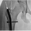
The center of femoral head to trochanteric tip vertical offset is normally zero. This affords maximum efficiency for the abductors. In preadolescent children, this distance is not measurable on plain radiographs because the tip of the trochanter is not ossified. In our series, transtrochanteric rod insertion did not result in any observed change in the center head/trochanteric distance (CH–TD)
Fig. 2
Fig. 2
a As a femur is rotated from orthogonal to 11° of (normal) version, and to simulated 30° of excessive anteversion, the shape of the head and neck, along with the apparent CH–TD appears to change. The projection artifact may lead to spurious conclusions regarding the femoral head–neck offset. b The projected femoral neck-shaft is altered as a result of positioning, causing parallax distortion. This is why it is not possible to accurately document the neck-shaft angle on plain AP images. It would also lead the unwary to conclude that, by correcting anteversion with an antegrade intramedullary rod, there is an iatrogenic change in the neck-shaft angle. Therefore, we chose not to measure the neck-shaft angle in this series
Fig. 3
Fig. 3
The new generation of trochanteric-entry, antegrade intramedullary femoral rods mitigates against iatrogenic osseous necrosis that had been previously reported with straight, piriformis entry rods. Presumably, the distance from the circumflex vessels protects against physical or thermal trauma during reaming
Fig. 4
Fig. 4
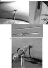
a Through a 1-cm incision, a stout drill bit (dotted oval), with a self-centering tip and short flutes, is employed to drill several transverse holes to mark the osteotomy site and decompress the femur for reaming. b These holes are connected with a ½-inch Ilizarov wrench (hexagonal handle) that is then torqued with a wrench. Advance the guide pin and rod past the osteotomy site before correcting the anteversion. Insert the distal interlocking screw first (freehand), then the proximal (jig)
Fig. 5
Fig. 5
The core diameter of 3.0 mm for the screw has been increased to 3.7 mm for the bolt. None of the bolts have broken

References

    1. Gordon JE, Khanna N, Luhmann SJ, Dobbs MB, Ortman MR, Schoenecker PL. Intramedullary nailing of femoral fractures in children through the lateral aspect of the greater trochanter using a modified rigid humeral intramedullary nail: preliminary results of a new technique in 15 children. J Orthop Trauma. 2004;18(7):416–422. doi: 10.1097/00005131-200408000-00004. - DOI - PubMed
    1. Kanellopoulos AD, Yiannakopoulos CK, Soucacos PN. Closed, locked intramedullary nailing of pediatric femoral shaft fractures through the tip of the greater trochanter. J Trauma. 2006;60(1):217–222. doi: 10.1097/01.ta.0000199913.02341.d6. - DOI - PubMed
    1. Stevens P, Gilliland J, Anderson L, Mickelson J, Nielson J, Klatt J. Success of torsional correction after failed surgeries for patellofemoral pain and instability. Strat Trauma Limb Reconstr. 2013;9(1):5–12. doi: 10.1007/s11751-013-0181-8. - DOI - PMC - PubMed
    1. Kaneko M, Sakuraba K. Association between femoral anteversion and lower extremity posture upon single-leg landing: implications for anterior cruciate ligament surgery. J Phys Ther Sci. 2013;25:1213–1217. doi: 10.1589/jpts.25.1213. - DOI - PMC - PubMed
    1. Nguyen A, Schultz S, Schmitz R. Landing biomechanics in participants with different static lower extremity alignment profiles. J Athl Train. 2015;50(5):498–507. doi: 10.4085/1062-6050-49.6.03. - DOI - PMC - PubMed

LinkOut - more resources