. 2016 Jun 20:7:11914.
doi: 10.1038/ncomms11914.

KRT14 marks a subpopulation of bladder basal cells with pivotal role in regeneration and tumorigenesis

Affiliations

KRT14 marks a subpopulation of bladder basal cells with pivotal role in regeneration and tumorigenesis

George Papafotiou et al. Nat Commun. .

Abstract

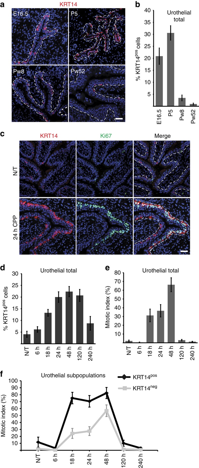
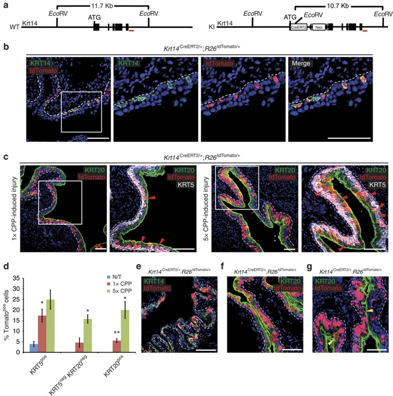
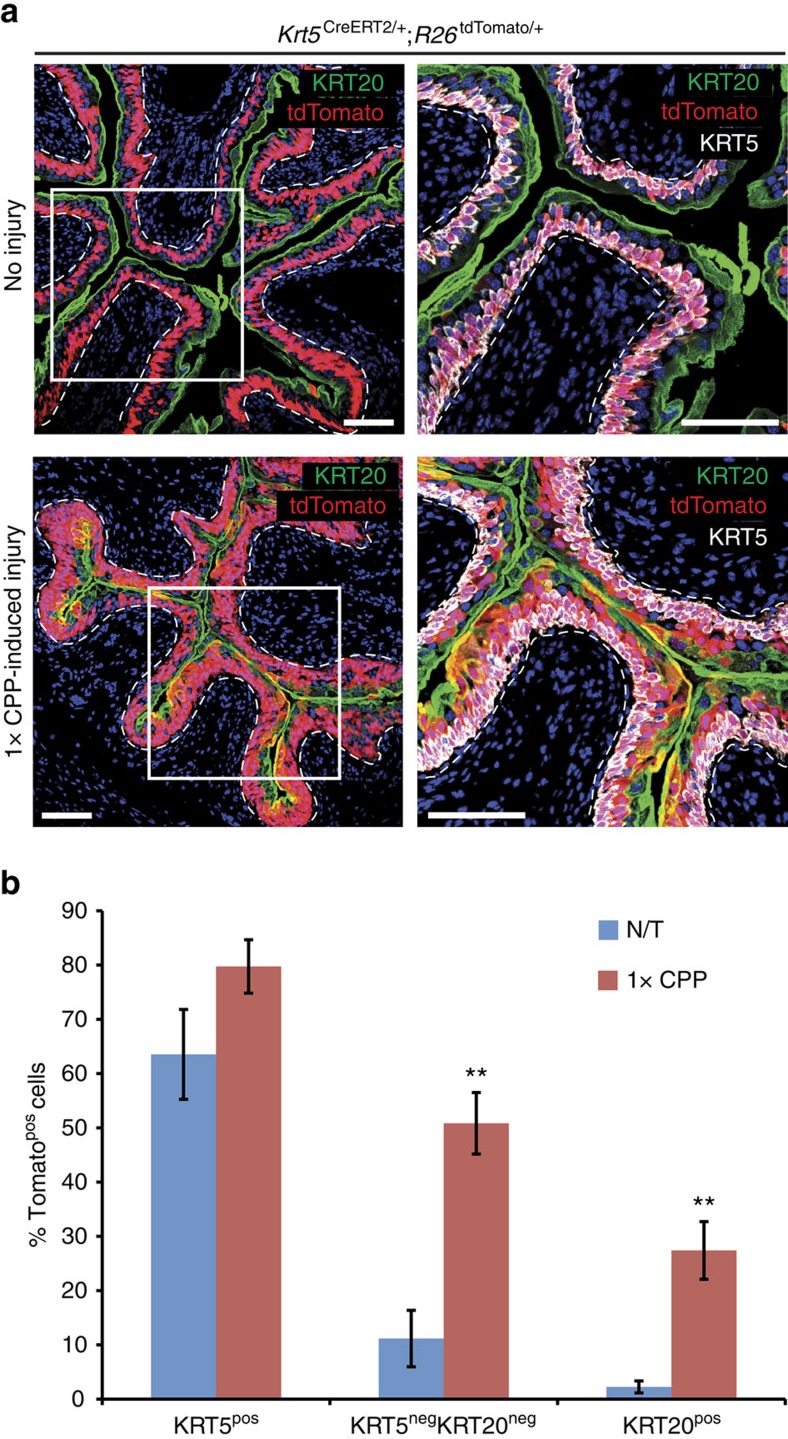
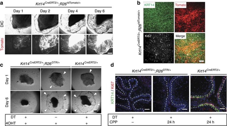
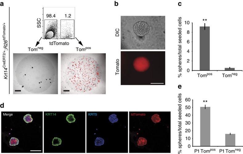
The urothelium is a specialized epithelium that lines the urinary tract. It consists of three different cell types, namely, basal, intermediate and superficial cells arranged in relatively distinct cell layers. Normally, quiescent, it regenerates fast upon injury, but the regeneration process is not fully understood. Although several reports have indicated the existence of progenitors, their identity and exact topology, as well as their role in key processes such as tissue regeneration and carcinogenesis have not been clarified. Here we show that a minor subpopulation of basal cells, characterized by the expression of keratin 14, possesses self-renewal capacity and also gives rise to all cell types of the urothelium during natural and injury-induced regeneration. Moreover, these cells represent cells of origin of urothelial cancer. Our findings support the hypothesis of basally located progenitors with profound roles in urothelial homoeostasis.


Figures

Figure 1
Figure 1. Dynamic changes in the number of KRT14pos cells in the normal development and tissue repair upon injury.
(a) Immunofluorescence (IF) against KRT14 on bladders from E16.5, P5, Pw8 and Pw52 bladders. (b) Quantification of KRT14pos cells at various developmental stages. Percentages are averages from three mice, and the total cells counted were 1,426, 1,669, 2,213 and 1,703 for E16.5, P5, Pw8 and Pw52, respectively. (c) IF against KRT14 and Ki67 on the bladder from an 8-week-old mouse before, and at 24 h after CPP injection. (d) Quantification of KRT14pos cells at 6, 18, 24, 48, 120 and 240 h post CPP injection. (e) Quantification of urothelial cell proliferation at the above time points post CPP injection. (f) Quantification of proliferation of KRT14pos and KRT14neg urothelial cells during CPP-induced injury and repair. Numbers in b, d and e are expressed as mean of percentages over total urothelial cells±s.e.m. Numbers in f are mean of percentages over KRT14pos or KRT14neg cells±s.e.m. For b, d, e and f, two group comparison was performed using the Student's t-test and the number of samples and P values can be found in the text, in Table 1 and in Supplementary Tables 1 and 2, respectively. For b, d and e, multiple comparison using Kruskal–Wallis test was also performed and P values were 0.0007, <0.0001 and 0.0001, respectively. Dash lines represent the basement membrane. Scale bars, 50 μm. N/T, not treated.
Figure 2
Figure 2. KRT14pos cells give rise to all urothelial lineages during both normal tissue turnover and chemical injury.
(a) Targeting strategy of the Krt14 locus with a CreERT2/Neo cassette. (b) Permanent labelling of KRT14+ basal urothelial cells by activation of a tdTomato transgene, by injecting an 8-week-old Krt14CreERT2/+;R26tdTomato/+ mouse with tamoxifen for five consecutive days and sacrificing 2 days later. (c) Sections of bladders of Krt14CreERT2/+;R26tdTomato/+ mice subjected to either one or five rounds of CPP-induced injury and full recovery. Red arrowheads point to Tomatopos/KRT5neg/low/KRT20neg intermediate cells. (d) Graph showing percentages of Tomatopos basal (KRT5pos), intermediate (KRT5negKRT20neg) and luminal (KRT20pos) cells in bladders from tamoxifen-injected, non CPP-treated Krt14CreERT2/+;R26tdTomato/+ mice or from Krt14CreERT2/+;R26tdTomato/+ mice after one or five rounds of CPP-induced injury and full recovery. Numbers are mean of percentages±s.e.m. Two group comparison was performed using the Student's t-test and the number of samples and P values can be found in Table 2. *P<0.05; **P<0.01. Multiple comparison using Kruskal–Wallis test was also performed and P values were 0.0107, 0.0014 and 0.0005 for Tomatopos basal (KRT5pos), intermediate (KRT5negKRT20neg) and luminal (KRT20pos) cell populations, respectively. (e) Sections of Krt14CreERT2/+;R26tdTomato/+ bladders from mice treated with tamoxifen at E16.5, and subsequently killed at either P5 without injury, or (f) after a round of CPP-induced injury and full recovery at Pw8. (g) Section of a bladder from a KRT14CreERT2/+;R26tdTomato/+ mouse killed at 10 months of age without injury. With the exception of embryonic labelling (e,f) the rest of the mice were injected with tamoxifen at the age of 8 weeks. Dash lines represent the basement membrane. Scale bars, 100 μm. N/T, not treated.
Figure 3
Figure 3. KRT5pos cells give rise to umbrella cells following CPP injury.
(a) Sections of bladders from Krt5CreERT2/+;R26tdTomato/+ mice with or without subsequent single round of CPP-induced injury and full recovery. Mice were injected with tamoxifen at the age of 8 weeks. Dash lines represent the basement membrane. Scale bars, 100 μm. (b) Graph showing percentages of Tomatopos basal (KRT5pos), intermediate (KRT5negKRT20neg) and luminal (KRT20pos) cells in bladders from tamoxifen-treated Krt5CreERT2/+;R26tdTomato/+ mice, either non CPP-treated or after a single round of CPP-induced injury and full recovery. Numbers are mean of percentages±s.e.m. For b, two group comparison was performed using the Student's t-test and the number of samples and P values can be found in Supplementary Table 3. **P<0.01. N/T, not treated.
Figure 4
Figure 4. KRT14pos cells support growth in ex vivo bladder tissue explant cultures.
(a) Urothelial explant cultures of a tamoxifen-injected Krt14CreERT2/+;R26tdTomato/+ 8-week-old mouse at 1, 2, 4 and 6 days. The tissue was obtained and cultured 2 days after the last tamoxifen injection. (b) KRT14 and Ki67 staining of the 6-day outgrowth of the explant shown in a. (c) Urothelial explant cultures of tissues obtained from tamoxifen-injected 8-week-old Krt14CreERT2/+;R26DTR/+ or Krt14CreERT2/+ mice, cultured either in the presence or absence of diphtheria toxin (DT). Four-hydroxytamoxifen (4OHT) was included in the culture medium for the first 12 h to ensure complete transgene recombination. (d) IF against KRT14 and Ki67 on bladders of experimental (Krt14CreERT2/+;R26tdTomato/+) and control (Krt14CreERT2/+) mice challenged with CPP and injected with DT. Scale bars, 1 mm (a,c); 100 μm (b,d).
Figure 5
Figure 5. KRT14pos cells show increased in vitro clonogenic capacity.
(a) Tomatoneg (Tomneg) and Tomatopos (Tompos) cells from tamoxifen-injected 8-week-old mice, were FACS sorted with Tomato positivity using a Becton Dickinson FACS Aria IIu cell sorter, and cultured in Matrigel. Cultures at 11 days are shown. (b) High magnification of a Tompos sphere from a. (c) Sphere-forming efficiency of Tompos (9.21±0.61%, n=10) and Tomneg (0.56±0.09%, n=10) urothelial cells from a. (d) IF against KRT5 and KRT14 of Matrigel-grown Tompos spheres indicating stratification. (e) Sphere forming efficiency of passaged (P1) Tompos (50.88±2.37%, n=10) and Tomneg (16.04±0.94%, n=9) urothelial cells obtained from Matrigel cultures from a. For c and e, two group comparison was performed using the Student's t-test. **P<0.0001. Scale bars represent 1 mm in a, and 100 μm in b and d.
Figure 6
Figure 6. Active Wnt/β-catenin signaling pathway is necessary for KRT14pos cell proliferation and bladder repair upon injury.
(a) In vivo treatment with 250 mg kg−1 indomethacin reduces CPP-induced proliferation (8.47±2.05% from 35.74±7.3% in vehicle-treated controls) and KRT14pos cell numbers (24 h after injury; 10.04±1.45% from 20.17±0.43% in vehicle-treated controls). Data are means of percentages ±s.e.m from four vehicle, or five Indomethacin-treated mice. Total cells counted were 5,108 and 9,947, respectively. (b) Clonogenic assays on Matrigel of total bladder populations cultured in the presence (421.6±10.8) or absence (705±48.4) of 100 μM indomethacin. Data are averages of five independent cultures. (c) Bladder explant cultures with increasing concentrations of indomethacin. (d) Axin 2 transcript levels (quantitative PCR data) in 3- day explants treated with 50 (56.1±2.7%) and 200 μM indomethacin (39.3±6.9%) plotted as percentage of Axin2 levels from vehicle-treated explants. (e) Clonogenic assays on Matrigel of bladder populations (10,000 cells) stably transduced with lentiviral vectors expressing a scrambled (167.38±7.14, n=8) or an anti-β-catenin (β-cat) shRNA (96.86±9.49, n=7). (f) Expression levels of β-catenin in 2-week total Matrigel cultures of primary bladder cells expressing anti-β-catenin (β-cat) shRNA (10.86±1.48%) plotted as percentage of β-cat levels from Matrigel cultures of primary bladder cells expressing scrambled shRNA. All data are mean values±s.e.m. For a, b and e, two group comparison was performed using the Student's t-test and *P<0.05; **P<0.01. Stars designate the bladder lumen.
Figure 7
Figure 7. KRT14pos cells are the cells of origin of bladder cancer.
(a) IF on bladders from untreated control 8-month-old mouse. (b) Widespread carcinoma in situ (CIS) in the bladder of a 6-month-old mouse treated with BBN for 4 months, showing remarkable KRT14pos cell population expansion, and extensive KRT14 and KRT5 colocalisation. (c) Invasive urothelial carcinoma (UC) in the bladder of an 8-month-old mouse treated with BBN for 6 months, showing extensive colocalisation of KRT14 and KRT5. (d-i) Krt14CreERT2/+;R26tdTomato/+ mice treated with tamoxifen before BBN treatment for 4 (d,f) or 6 months (e,g-i) showing variable histological abnormalities and cytokeratin profile. Stars designate the bladder lumen. Scale bars, 100 μm.

References

    1. Kreft M. E., Hudoklin S., Jezernik K. & Romih R. Formation and maintenance of blood-urine barrier in urothelium. Protoplasma 246, 3–14 (2010). - PubMed
    1. Farsund T. & Dahl E. Cell kinetics of mouse urinary bladder epithelium. III. A histologic and ultrastructural study of bladder epithelium during regeneration after a single dose of cyclophosphamide, with special reference to the mechanism by which polyploid cells are formed. Virchows Arch. B Cell Pathol. 26, 215–223 (1978). - PubMed
    1. Mysorekar I. U. Molecular regulation of urothelial renewal and host defenses during infection with uropathogenic Escherichia coli. J. Biol. Chem. 277, 7412–7419 (2002). - PubMed
    1. Shin K. et al. Hedgehog/Wnt feedback supports regenerative proliferation of epithelial stem cells in bladder. Nature 472, 110–114 (2011). - PMC - PubMed
    1. Gandhi D. et al. Retinoid signaling in progenitors controls specification and regeneration of the urothelium. Dev. Cell 26, 469–482 (2013). - PMC - PubMed

Publication types

MeSH terms