. 2016 Jun 20:7:11964.
doi: 10.1038/ncomms11964.

Development of background-free tame fluorescent probes for intracellular live cell imaging

Affiliations

Development of background-free tame fluorescent probes for intracellular live cell imaging

Samira Husen Alamudi et al. Nat Commun. .

Abstract

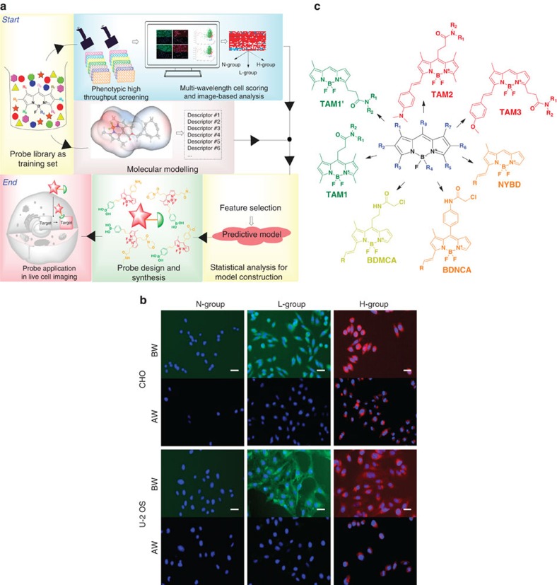
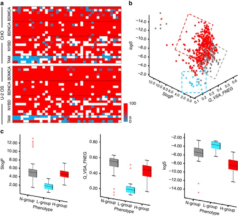
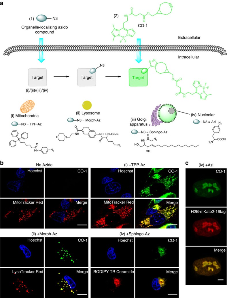
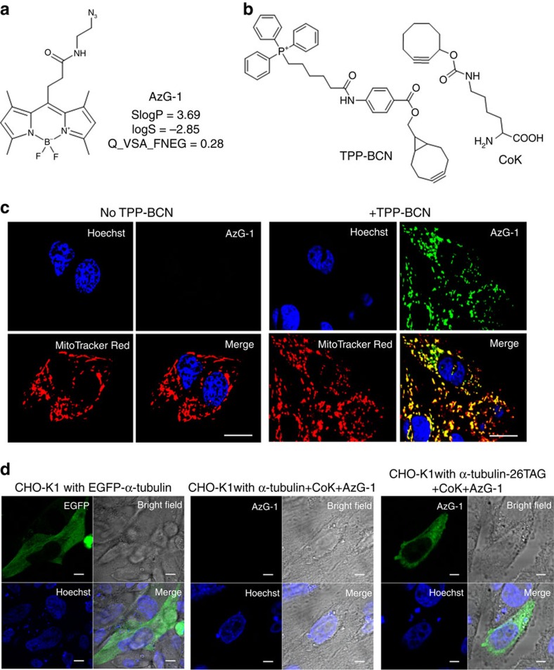
Fluorescence labelling of an intracellular biomolecule in native living cells is a powerful strategy to achieve in-depth understanding of the biomolecule's roles and functions. Besides being nontoxic and specific, desirable labelling probes should be highly cell permeable without nonspecific interactions with other cellular components to warrant high signal-to-noise ratio. While it is critical, rational design for such probes is tricky. Here we report the first predictive model for cell permeable background-free probe development through optimized lipophilicity, water solubility and charged van der Waals surface area. The model was developed by utilizing high-throughput screening in combination with cheminformatics. We demonstrate its reliability by developing CO-1 and AzG-1, a cyclooctyne- and azide-containing BODIPY probe, respectively, which specifically label intracellular target organelles and engineered proteins with minimum background. The results provide an efficient strategy for development of background-free probes, referred to as 'tame' probes, and novel tools for live cell intracellular imaging.


Conflict of interest statement

Y.-T.C., S.H.A. and R.S. are the inventors of CO-1 and AzG-1 for which a patent has been applied The remaining authors declare no competing financial interests.

Figures

Figure 1
Figure 1. Experimental set and probes' cellular responses.
(a) Flowchart of the overall experimental strategy. (b) Representative cellular responses in CHO and U-2 OS. Cells were stained with probes at 1 μM final concentration. The overlayed images of Hoechst33324 (blue) and probe signals (according to emission wavelength of the probes) show three phenotypic groups: cell-impermeable group where probes are unable to stain the cell (N-group); cell permeable with low nonspecific binding group (L-group); and cell permeable with high nonspecific binding group (H-group). BW and AW indicate image before and after washing, respectively. Probes signal is significantly decreased after washing in L-group, while is retained in H-group. Scale bar, 10 μm (green, red and blue signals are from FITC, Texas Red and DAPI channel, respectively). (c) Core structures of the BODIPY libraries used in training set.
Figure 2
Figure 2. Predictive model.
(a) Heat maps of RR for each library in U-2 OS and CHO cell lines. White colour represents probes in N-group. The red and torquise colour represents high and low RR, respectively. (b) Three dimensional scatter plot for three key descriptors: SlogP, Q_VSA_FNEG and logS. Grey, turquoise and red dotted box represents area containing mostly probes from N-group, L-group and H-group, respectively. (c) Preferred criteria for ‘tame' probes. Descriptor values in each group are presented as box plots with medians, quartiles, and interquartile range. The whiskers represent the minimum and maximum scores, and the red asterisks show extreme scores.
Figure 3
Figure 3. Live cell imaging with CO-1.
(a) Chemical structures and schematic illustration of the covalent labelling of azide-tagged organelles using CO-1 in live cells. (b) Fluorescence imaging of mitochondria, lysosome and golgi apparatus in U-2 OS cells labelled with CO-1. Cells pre-treated with TPP-Az, Morph-Az or Sphingo-Az were incubated with 2 μM CO-1 in growth media at 37 °C for 1 h and followed by counterstaining with organelle trackers. Scale bar, 15 μm. (c) Fluorescence imaging of histone H2B in live U-2 OS. U-2 OS cells were co-transfected with plasmids pIre-Azi3 and pmH2B-6-mKate2-16tag to incorporate UAA Azi into H2B-mKate2 at site 16. After Azi incorporation, cells were labelled with 10 μM CO-1 for 90 min at 37 °C. Cells were then washed and imaged. The mKate2 (red) signals were detected in cell nuclei only, and colocalized with the CO-1 (green) signals in the merged image. Scale bar, 5 μm.
Figure 4
Figure 4. Live cell imaging with AzG-1
(a) Chemical structures of azide-bearing probe AzG-1 and its descriptor values. (b) Structure of triphenylphosphonium analogues TPP-BCN and cyclooctyne-containing unnatural amino acid, CoK. (c) Fluorescence imaging of mitochondria in U-2 OS cells labelled with AzG-1. Cells were incubated without (left side) and with (right side) TPP-BCN and then labelled with 10 μM AzG-1 for 2 h followed by counterstaining with mitochondria marker. Scale bar, 10 μm. (d) Fluorescence imaging of α-tubulin in live CHO-K1 cells. CHO-K1 cells were co-transfected with plasmids pCoKRS-tRNA and pTub-26TAG to incorporate CoK into α-tubulin at position 26. After incorporation of CoK, cells were labelled with 10 μM AzG-1 for 2 h at 37 °C. CHO-K1 cells were also transfected with plasmid pTubwt to express wild type α-tubulin and treated with CoK and AzG-1 in the same way. CHO-K1 cells transfected with plasmid pEGFP-Tubwt expressing EGFP-fused wild type α-tubulin was used as a control. Live cell images show that AzG-1 labelled mitochondria (c) and is conjugated specifically with CoK-bearing α-tubulin (d). Scale bar, 10 μm.

References

    1. Jung D., Min K., Jung J., Jang W. & Kwon Y. Chemical biology-based approaches on fluorescent labeling of proteins in live cells. Mol. BioSyst. 9, 862–872 (2013). - PubMed
    1. Baskin J. M. Copper-free click chemistry for dynamic in vivo imaging. Proc. Natl Acad. Sci. USA 104, 16793–16797 (2007). - PMC - PubMed
    1. Agard N. J., Prescher J. A. & Bertozzi C. R. A strain-promoted [3+2] azide-alkyne cycloaddition for covalent modification of biomolecules in living systems. J. Am. Chem. Soc. 126, 15046–15047 (2004). - PubMed
    1. Beatty K. E., Szychowski J., Fisk J. D. & Tirrel D. A. A BODIPY-cyclooctyne for protein imaging in live cells. Chembiochem 12, 2137–2139 (2011). - PMC - PubMed
    1. Fernandez-Suarez M. et al. Redirecting lipoic acid ligase for cell surface protein labeling with small-molecule probes. Nat. Biotech. 25, 1483–1487 (2007). - PMC - PubMed

Publication types

MeSH terms