

Mitochondrial function in hypoxic ischemic injury and influence of aging
- PMID: 27321753
- PMCID: PMC5161736
- DOI: 10.1016/j.pneurobio.2016.06.006
Mitochondrial function in hypoxic ischemic injury and influence of aging
Abstract
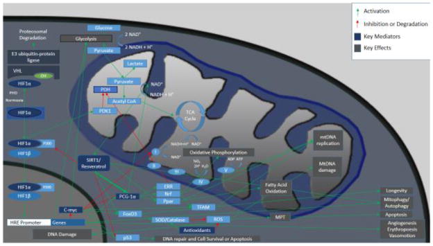
Mitochondria are a major target in hypoxic/ischemic injury. Mitochondrial impairment increases with age leading to dysregulation of molecular pathways linked to mitochondria. The perturbation of mitochondrial homeostasis and cellular energetics worsens outcome following hypoxic-ischemic insults in elderly individuals. In response to acute injury conditions, cellular machinery relies on rapid adaptations by modulating posttranslational modifications. Therefore, post-translational regulation of molecular mediators such as hypoxia-inducible factor 1α (HIF-1α), peroxisome proliferator-activated receptor γ coactivator α (PGC-1α), c-MYC, SIRT1 and AMPK play a critical role in the control of the glycolytic-mitochondrial energy axis in response to hypoxic-ischemic conditions. The deficiency of oxygen and nutrients leads to decreased energetic reliance on mitochondria, promoting glycolysis. The combination of pseudohypoxia, declining autophagy, and dysregulation of stress responses with aging adds to impaired host response to hypoxic-ischemic injury. Furthermore, intermitochondrial signal propagation and tissue wide oscillations in mitochondrial metabolism in response to oxidative stress are emerging as vital to cellular energetics. Recently reported intercellular transport of mitochondria through tunneling nanotubes also play a role in the response to and treatments for ischemic injury. In this review we attempt to provide an overview of some of the molecular mechanisms and potential therapies involved in the alteration of cellular energetics with aging and injury with a neurobiological perspective.
Keywords: Alzheimer’s disease; Apoptosis; Autophagy; Blood brain barrier; Hypoxia; Intermitochondrial signal propagation; Ischemia/reperfusion; Mitokine; Mitoquinone; Nuclear-mitochondria cross-talk; Oxidative phosphorylation; Parkinson’s disease; Pseudohypoxia; Resveratrol, SIRT1, sirtuins; Stroke, myocardial infarction; Tempol; Tunneling nanotube.
Copyright © 2016 Elsevier Ltd. All rights reserved.
Figures

References
-
- Adam T, Sharp S, Opie LH, Lecour S. Loss of cardioprotection with ischemic preconditioning in aging hearts: role of sirtuin 1? J Cardiovasc Pharmacol Ther. 2013;18:46–53. - PubMed
-
- Adlam VJ, Harrison JC, Porteous CM, James AM, Smith RA, Murphy MP, Sammut IA. Targeting an antioxidant to mitochondria decreases cardiac ischemia-reperfusion injury. FASEB J. 2005;19:1088–1095. - PubMed
-
- Adler A, Messina E, Sherman B, Wang Z, Huang H, Linke A, Hintze TH. NAD(P)H oxidase-generated superoxide anion accounts for reduced control of myocardial O2 consumption by NO in old Fischer 344 rats. Am J Physiol Heart Circ Physiol. 2003;285:H1015–1022. - PubMed
-
- Ahmad T, Mukherjee S, Pattnaik B, Kumar M, Singh S, Kumar M, Rehman R, Tiwari BK, Jha KA, Barhanpurkar AP, Wani MR, Roy SS, Mabalirajan U, Ghosh B, Agrawal A. Miro1 regulates intercellular mitochondrial transport & enhances mesenchymal stem cell rescue efficacy. EMBO J. 2014;33:994–1010. - PMC - PubMed
Publication types
MeSH terms
Grants and funding
LinkOut - more resources
Full Text Sources
Other Literature Sources
Medical
