Review
. 2016 Jun 18;21(6):797.
doi: 10.3390/molecules21060797.

Capsaicin, Nociception and Pain

Affiliations
Review

Capsaicin, Nociception and Pain

Bárbara Frias et al. Molecules. .

Abstract

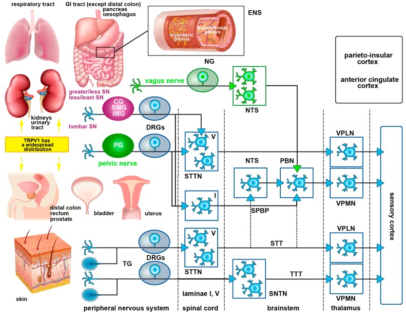
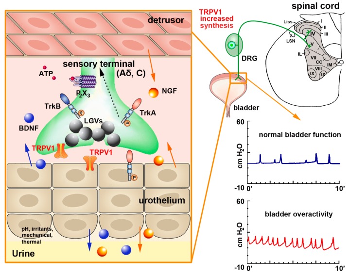
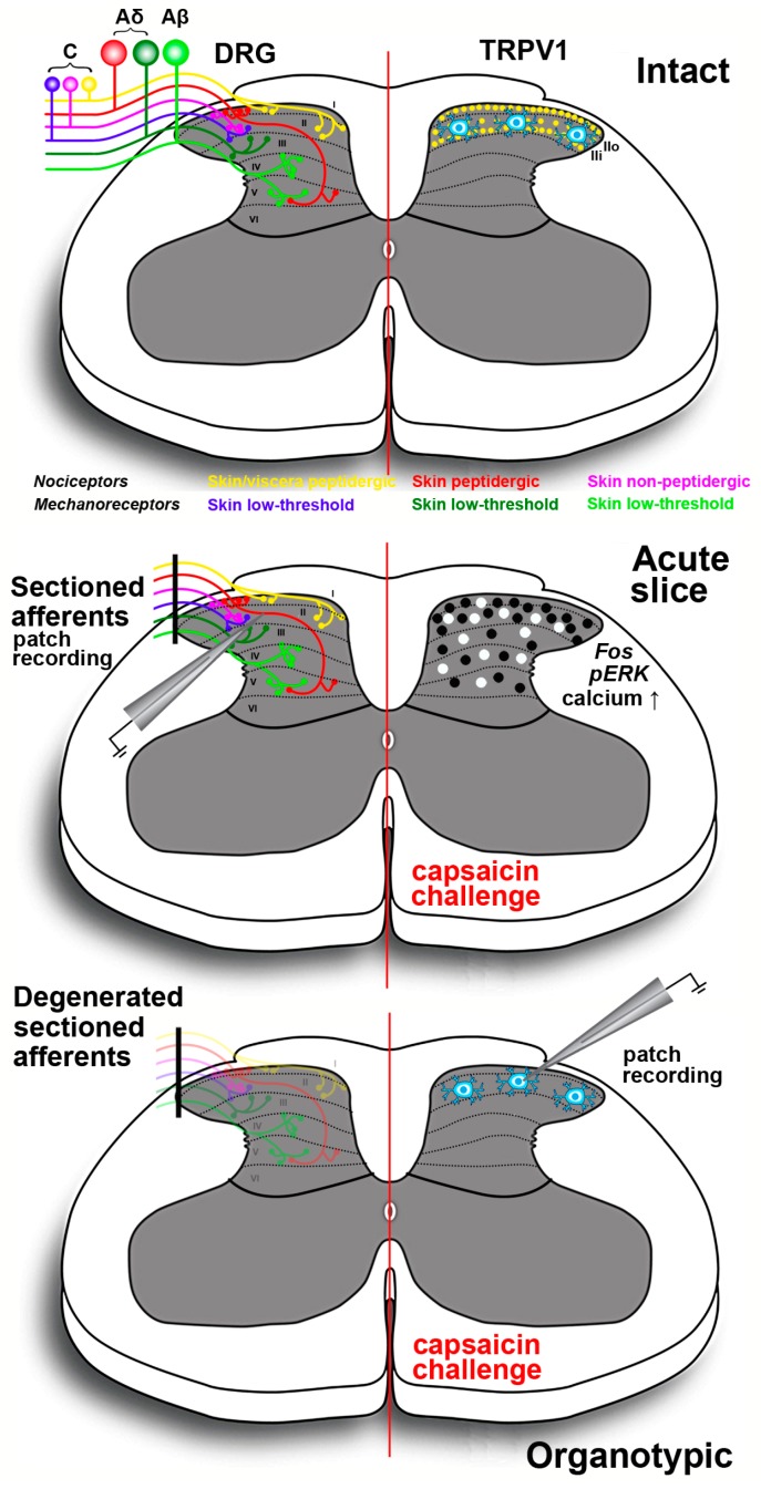
Capsaicin, the pungent ingredient of the hot chili pepper, is known to act on the transient receptor potential cation channel vanilloid subfamily member 1 (TRPV1). TRPV1 is involved in somatic and visceral peripheral inflammation, in the modulation of nociceptive inputs to spinal cord and brain stem centers, as well as the integration of diverse painful stimuli. In this review, we first describe the chemical and pharmacological properties of capsaicin and its derivatives in relation to their analgesic properties. We then consider the biochemical and functional characteristics of TRPV1, focusing on its distribution and biological effects within the somatosensory and viscerosensory nociceptive systems. Finally, we discuss the use of capsaicin as an agonist of TRPV1 to model acute inflammation in slices and other ex vivo preparations.

Keywords: TRPV1 receptor; analgesia; capsaicin; nociception; resinferatoxin; sensitization; somatic pain; vanilloids; visceral pain.


Conflict of interest statement

The authors declare no conflict of interest.

Figures

Figure 1
Figure 1
Illustration depicting TRPV1 receptor distribution in body organs and the main visceral and somatic pain pathways. TRPV1 is found in several body organs and when its expression increases, it contributes to the development of visceral and somatic pain. Afferent fibers innervating the viscera project to the CNS following the course of autonomic sympathetic and parasympathetic nerves. Afferent sympathetic fibers originating from the thoracolumbar DRGs follow the hypogastric, lumbar and splanchnic nerves (SN) after traversing the sympathetic prevertebral (celiac ganglion—CG, superior mesenteric ganglion- SMG and inferior mesenteric ganglia—IMG) and paravertebral ganglia (in magenta). Ascending projections from lamina I neurons in the spinal cord travel along the spinoparabrachial pathway (SPBP) to the parabrachial nucleus (PBN), whereas projections from deep dorsal horn neurons (STTN) travel along the spinothalamic tract (STT) to thalamic nuclei (VPMN and VPLN). The parasympathetic sensory innervation (in green) follows the vagus and pelvic nerve that terminate in the brainstem and lumbosacral cord, respectively. The gastrointestinal tract also has its own autonomic intrinsic nervous system—the enteric nervous system (ENS) — constituted by the submucosal (Meissner’s) and myenteric (Auerbach’s) plexuses. Enteric plexuses play a key role for communication with the autonomic extrinsic nervous system in several GI functions. Nociceptive somatic inputs from all the parts of the body, except the head, are transmitted to spinothalamic projection neurons in the dorsal horn of the spinal cord. These neurons in turn, reach the neurons in the ventro-postero lateral nucleus of the thalamus (VPLN) through the STT. Nociceptive somatic inputs (for simplicity, only the skin is depicted but these inputs also derive from the muscles, tendons, bones and joints) from the head are relayed to the spinal nucleus of the trigeminal nerve (SNTN) and then, along the trigeminothalamic fibers to the ventro-postero medial nucleus of the thalamus (VPMN). Finally, nociceptive input is transferred to the sensory cortex where it is perceived as pain. Affective, emotional and autonomic aspects of pain are processed in other cortical areas (black).
Figure 2
Figure 2
Schematics of TRPV1 localization and function in the urinary bladder and its contribution to bladder dysfunction. In the urinary bladder, TRPV1 can be found in sensory afferents and in the urothelium. Upon mechanical injury or inflammation TRPV1 levels are increased together with those of substance P and CGRP (stored in large granular vesicles—LGVs—large gray spheres) Nerve growth factor (NGF—orange spheres) is also released by the detrusor smooth muscle and the urothelium (orange arrows). NGF activates tropomyosin-related kinase A (TrkA) receptors expressed on afferent terminals, contributing to sensitization of neuronal TRPV1. The TrkA-NGF complex is internalized and retrogradely transported (dashed line) to neurons in lumbosacral dorsal root ganglia (DRGs), where de novo transcription of TRPV1 and additional sensory ion channels (including purinergic P2X3 receptor for ATP—small red spheres) is initiated. These newly synthesized ion channels are anterogradely transported back to afferent terminals to contribute to peripheral hypersensitivity. The urothelium also potentially produces brain-derived nerve factor (BDNF—blue spheres), which binds to tropomyosin-related kinase B (TrkB) receptors, further contributing to sensitization. This also participates to the development of bladder over activity, as shown by the increase number of bladder contractions in the graph at bottom right of the figure.
Figure 3
Figure 3
Schematic drawing of the spinal cord slices preparations exploiting the agonist action of capsaicin onto TRPV1 expressing neurons and fibers in the dorsal horn. Top panel shows at left the laminar distribution of primary afferent terminals originating from different classes of nociceptors in DRG neurons; at right the distribution of TRPV1 in peptidergic terminals (yellow dots) originating from skin and viscera in laminae I-II. Receptors are less densely localized in the outer part of lamina II (IIo) as compared to lamina I and the inner part of lamina II (IIi). Lamina II also contains TRPV1-IR interneurons that are spared in organotypically cultured slices (bottom panel). For simplicity, proprioceptive muscle afferents are not represented. In acute slices (middle panel) primary afferents are severed at dorsal roots but their terminals remain functional for several hours allowing performing electrophysiological recording, calcium imaging and proto-oncogene c-Fos/pERK immunocytochemistry. The black dots indicate the distribution of proto-oncogene c-Fos/pERK-IR neurons after capsaicin challenge. White dots exemplify the location of the cells responding to the vanilloid with increased intracellular calcium concentration in real-time confocal imaging. In organotypic cultures (bottom panel) sectioned primary afferents degenerate and thus, only the TRPV1-IR lamina II neurons are spared. This type of preparation is useful to isolate the effects of TRPV1 activation on these neurons, in the absence of primary afferent input.

References

    1. Thresh J.C. Isolation of capsacin. Pharm. J. Tr. 1876;6:941–947.
    1. Nelson E.K. The constitution of capsaicin, the pungent principle of capsicum. II. J. Am. Chem. Soc. 1920;42:597–599.
    1. De A.K., editor. Capsicum: The Genus Capsicum. CRC Press; London, UK: 2003.
    1. Musfiroh I., Mutakin M., Angelina T., Muchtaridi M. Capsaicin level of various capsicum fruits. Int. J. Pharm. Pharm. Sci. 2013;5:248–251.
    1. Caterina M.J., Schumacher M.A., Tominaga M., Rosen T.A., Levine J.D., Julius D. The capsaicin receptor: A heat-activated ion channel in the pain pathway. Nature. 1997;389:816–824. - PubMed