. 2016 Jun 20;11(6):e0157586.
doi: 10.1371/journal.pone.0157586. eCollection 2016.

The Barley Powdery Mildew Effector Candidates CSEP0081 and CSEP0254 Promote Fungal Infection Success

Affiliations

The Barley Powdery Mildew Effector Candidates CSEP0081 and CSEP0254 Promote Fungal Infection Success

Ali Abdurehim Ahmed et al. PLoS One. .

Abstract

Effectors play significant roles in the success of pathogens. Recent advances in genome sequencing have revealed arrays of effectors and effector candidates from a wide range of plant pathogens. Yet, the vast majority of them remain uncharacterized. Among the ~500 Candidate Secreted Effector Proteins (CSEPs) predicted from the barley powdery mildew fungal genome, only a few have been studied and shown to have a function in virulence. Here, we provide evidence that CSEP0081 and CSEP0254 contribute to infection by the fungus. This was studied using Host-Induced Gene Silencing (HIGS), where independent silencing of the transcripts for these CSEPs significantly reduced the fungal penetration and haustoria formation rate. Both CSEPs are likely required during and after the formation of haustoria, in which their transcripts were found to be differentially expressed, rather than in epiphytic tissue. When expressed in barley leaf epidermal cells, both CSEPs appears to move freely between the cytosol and the nucleus, suggesting that their host targets locate in these cellular compartments. Collectively, our data suggest that, in addition to the previously reported effectors, the barley powdery mildew fungus utilizes these two CSEPs as virulence factors to enhance infection.


Conflict of interest statement

Competing Interests: The authors have declared that no competing interests exist.

Figures

Fig 1
Fig 1. HIGS-mediated silencing of CSEP0081 and CSEP0254 decreased Bgh haustorial formation success.
Barley leaves, bombarded with RNAi and GUS reporter constructs, were infected with Bgh and scored for fungal haustoria formation. The haustorial formation rate was calculated as the ratio of haustoria-containing transformed cells (GUS expressing cells) divided by the total number of transformed cells. Relative haustorial formation rate was computed relative to the empty vector control of each experiment, which was set to 100%. Data shown are mean values of five independent experiments, but CSEP0145, CSEP0216, CSEP0222, CSEP0398 and Mlo were excluded from two of those. All values are presented ± SE. ** refers to significant differences compared to the empty vector control (P < 0.01). Arrow indicates haustoria. Scale bar, 20 μm.
Fig 2
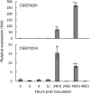
Fig 2. Relative expression levels of CSEP0081 and CSEP0254 during Bgh infection.
The CSEP transcripts were monitored using RT-qPCR. Total RNA was isolated from Bgh-infected barley leaves. Haustorial and epiphytic expression was analysed separately at 24 and 48 hpi (H and E, respectively). Expression of Bgh glyceraldehyde 3-phosphate dehydrogenase (GAPDH) was used to normalize the CSEPs expression in each sample. Expression was determined relative to 0 hpi, arbitrarily set to one. Data represent means of three independent biological repeats, each with two technical repeats, ± SE. Statistical analysis of biological repeats: t test relative to 0 hpi; **, P<0.01; ***, P<0.001.
Fig 3
Fig 3. CSEP0081 fused to YFP localizes to the cytosol and the nucleus of barley leaf epidermal cells.
Construct encoding CSEP0081, lacking signal peptide, with YFP fused to its C-terminus was co-expressed with a free mCherry marker construct in barley leaf epidermal cells using particle bombardment. Localization was monitored 1 to 3 days later. The CSEP0081-YFP fusion proteins co-localized with mCherry in the cytosol and the nucleus, shown by a complete merge of yellow and red signals. C, cytosol. N, nucleus. Scale bar, 20 μm.

References

    1. Cook DE, Mesarich CH, Thomma BP. Understanding plant immunity as a surveillance system to detect invasion. Annu Rev Phytopathol. 2015;53: 541–563. 10.1146/annurev-phyto-080614-120114 - DOI - PubMed
    1. Stuart LM, Paquette N, Boyer L. Effector-triggered versus pattern-triggered immunity: how animals sense pathogens. Nat Rev Immunol. 2013;13: 199–206. 10.1038/nri3398 - DOI - PMC - PubMed
    1. Jones JDG, Dangl JL. The plant immune system. Nature. 2006;444: 323–329. - PubMed
    1. de Wit PJGM. How plants recognize pathogens and defend themselves. Cell Mol Life Sci. 2007;64: 2726–2732. - PMC - PubMed
    1. Stergiopoulos I, de Wit P. Fungal effector proteins. Annu Rev Phytopathol 2009;47:233–263. 10.1146/annurev.phyto.112408.132637 - DOI - PubMed