. 2016 Aug 9;7(32):51082-51095.
doi: 10.18632/oncotarget.9013.

The anti-apoptotic PON2 protein is Wnt/β-catenin-regulated and correlates with radiotherapy resistance in OSCC patients

Affiliations

The anti-apoptotic PON2 protein is Wnt/β-catenin-regulated and correlates with radiotherapy resistance in OSCC patients

Maximilian Krüger et al. Oncotarget. .

Abstract

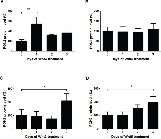
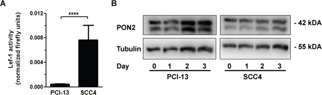
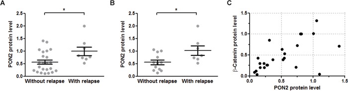
Aberrant Wnt signaling and control of anti-apoptotic mechanisms are pivotal features in different types of cancer to undergo cell death programs. The intracellular human enzyme Paraoxonase-2 (PON2) is known to have anti-apoptotic properties in leukemia and oral squamous cell cancer (OSCC) cells. However, the distinct regulating pathways are poorly understood. First, we present a so far unknown regulation of PON2 protein expression through the Wnt/GSK3β/β-catenin pathway in leukemia and OSCC cells. This was confirmed via in silico analysis, promoter reporter studies and treatment of multiple cell lines (K562, SCC-4, PCI-13) with different Wnt ligands/inhibitors in vitro. Ex vivo analysis of OSCC patients revealed a correlation between PON2 and β-catenin expression in tumor tissue. Higher PON2 expression in OSCC is associated with relapse independently of treatment (e.g. surgery/radio-/chemotherapy). These results emphasize the clinical impact of the newly described regulation of PON2 through Wnt/GSK3β/β-catenin. More importantly, the study revealed the fundamental finding of an overall Wnt/GSK3β/β-catenin dependent regulation of PON2 in different cancers, which was confirmed by systematic and multimethodological approaches. Thus, the herein presented mechanistic insight contributes to a better understanding of tumor specific escape from cell death strategies and suggests PON2 as a new potential biomarker for therapy resistance or as a prognostic tumor marker.

Keywords: Wnt / beta-catenin; leukemia; oral squamous cancer cell; paraoxonase-2; tumor.


Conflict of interest statement

The authors declare no conflicts of interest.

Figures

Figure 1
Figure 1. PON2 is highly overexpressed in Imatinib resistant cells, but neither Imatinib nor ERK have a direct effect on the expression
A. Lama84 and KCL22 cells sensitive (S) or resistant (R) to Imatinib were analyzed for PON2 mRNA levels by qRT-PCR. B. K562 cells were treated with Imatinib (0.3 μM) for indicated times. Lysates were analyzed by Western blotting with anti-PON2 and anti–α-tubulin antibodies. C. Similar analysis as in B but employing the ERK inhibitor PD 98059 (10 μM). Symbols represent mean ± S.E.M. n = 3 – 13; *P < 0.05.
Figure 2
Figure 2. Identification of pathways and / or transcription factors involved in PON2 regulation
A. The indicated three databases were used for in silico prediction of transcription factors (TFs) that bind to a 10-kbp sequence putatively containing PON2-regulating elements upstream transcription start. See text for further details. B. Scheme depicting evolutionary conserved regions in human (h) and mouse (m) genomic context upstream PON2 transcription site. Peaks indicate numbers of conserved (above 50%) nucleotides; blue = coding exons; yellow = UTRs; green = transposable elements; red = conserved areas in intergenic regions; R1 – R4 with bars = evolutionary conserved regions; asterisks = Lef-1 sites; blue line with arrows within = protein-coding region. C. Control DNA or a 7,5-kbp stretch of human genomic DNA upstream PON2 transcription start was bound to beads and incubated with nuclear extracts isolated from endothelial EA.hy 926 or K562 cells (±SB216763). TFs binding to PON2 promoter but not to control DNA were identified by mass spectrometry employing three independent biological repeats. The graph depicts binding of SMARCA family members.
Figure 3
Figure 3. Inhibition of GSK-3β results in up-regulation of PON2 expression in K562 cells
K562 cells were treated with valproic acid (1mM) for the indicated times and analyzed for A. PON2 mRNA levels by qRT-PCR or B. PON2 protein levels by Western blotting. C. K562 cells were transfected with a PON2 promoter activity reporter plasmid and were untreated or treated with the GSK-β inhibitor SB216763 (25μM) for the indicated times. Subsequently PON2 promoter induction was quantified. D, E. K562 cells were treated with SB216763 (25μM) for indicated times and quantified for PON2 mRNA (D) or PON2 and β-catenin protein level (E). One representative blot is shown (right). Symbols represent mean ± S.E.M. n = 4; * P < 0.05; **P < 0.01; ***P < 0.001.
Figure 4
Figure 4. PON2 expression is regulated by LEF-1 and TCF4, but not TCF1
K562 cells were co-transfected with plasmids encoding a firefly luciferase gene under the expressional control of the 7-LEF-fos-luc A. or PON2 promoter fragment B., a plasmid for constitutive expression of Renilla luciferase (normalization) and pEGFP-C1, pEGFP-C1-dnLEF-1, pEGFP-C1-TCF1 or pEGFP-C1-TCF4 plasmids. At 4 h after transfection, cells were treated with SB216763 (25 μM) for 24 h. Subsequently 7-LEF-fos-luc or PON2 promoter induction was analyzed, normalized to Renilla luciferase activity and expressed as fold induction. Symbols represent mean ± S.E.M. n = 2; * P < 0.05; **P < 0.01; ***P < 0.001.
Figure 5
Figure 5. The Wnt ligands 3a and 5a induce PON2 expression
A-B. Quantitative analysis of PON2 protein expression in K562 cells after treatment with recombinant Wnt3a (100 ng/ml) or Wnt5a (200 ng/ml). PON2 expression was normalized to GAPDH. C-D. The same analysis as in A + B, but using primary human umbilical vein endothelial cells (HUVECs). Symbols represent mean ± S.E.M. n = 2 – 5; *P < 0.05; **P < 0.01.
Figure 6
Figure 6. GSK-3β inhibition upregulated PON2 only in oral squamous cancer cell lines with low LEF-1 activity
A. Oral squamous carcinoma cell lines SCC4 and PCI-13 were transfected with a LEF-1 activity gene reporter plasmid. At 24 h after transfection, LEF-1 promoter induction was analyzed, normalized to Renilla luciferase activity and expressed as fold induction. B. PCI-13 and SCC4 cells were treated with SB216763 (25μM) for indicated times and analyzed for PON2 and a-tubulin protein levels by Western blotting. Blots are representative of three others. Symbols represent mean ± S.E.M. n = 3; ****P < 0.0001 (t-test).
Figure 7
Figure 7. PON2 expression associated with relapse occurrence and β-catenin levels in oral squamous cell tumors
A. Benign mucosa and tumors were isolated from 32 OSCC patients during tumor-removing surgery and analyzed for PON2 protein level by Western blotting. Patients were grouped as to whether they experienced a relapse during the observation period or not. B. Subgroup analysis of the patients from panel A analyzing only those patients that received radio-/chemotherapy after surgical tumor removal. C. Subgroup analysis of 24 patients (with or without radio-/chemotherapy) for PON2 and β-catenin protein levels in named samples (tumor / benign mucosa). PON2 and β-catenin expression correlated with Spearman r = 0.8313; 95% CI = 0.6363-0.9265; p<0.0001; Pearson r = 0.7693; 95% CI = 0.5304-0.895; p<0.0001.

References

    1. Siegel RL, Miller KD, Jemal A. Cancer statistics, 2015. CA Cancer J Clin. 2015;65:5–29. - PubMed
    1. Hanahan D, Weinberg RA. Hallmarks of cancer: The next generation. Cell. 2011;144:646–674. - PubMed
    1. Logan CY, Nusse R. The wnt signaling pathway in development and disease. Annu Rev Cell Dev Biol. 2004;20:781–810. - PubMed
    1. Morgan RG, Ridsdale J, Tonks A, Darley RL. Factors affecting the nuclear localization of beta-catenin in normal and malignant tissue. J Cell Biochem. 2014;115:1351–1361. - PubMed
    1. Staal FJ, Clevers HC. Wnt signalling and haematopoiesis: A wnt-wnt situation. Nat Rev Immunol. 2005;5:21–30. - PubMed

MeSH terms