

Focal liver hyperplasia in a patient with Alagille syndrome: Diagnostic difficulties. A case report
- PMID: 27322896
- PMCID: PMC4916051
- DOI: 10.1016/j.ijscr.2016.03.032
Focal liver hyperplasia in a patient with Alagille syndrome: Diagnostic difficulties. A case report
Abstract
Introduction: Alagille syndrome is a multisystem autosomal disorder. The main clinical features are chronic cholestasis due to paucity of intrahepatic bile ducts, which can progress to cirrhosis and liver failure.
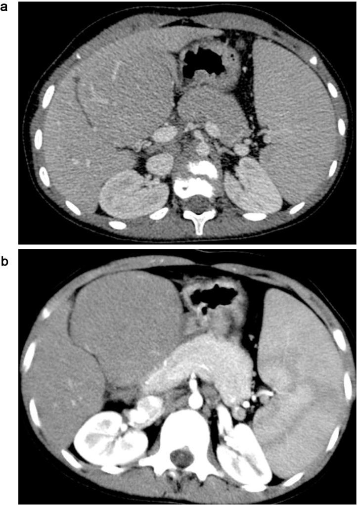
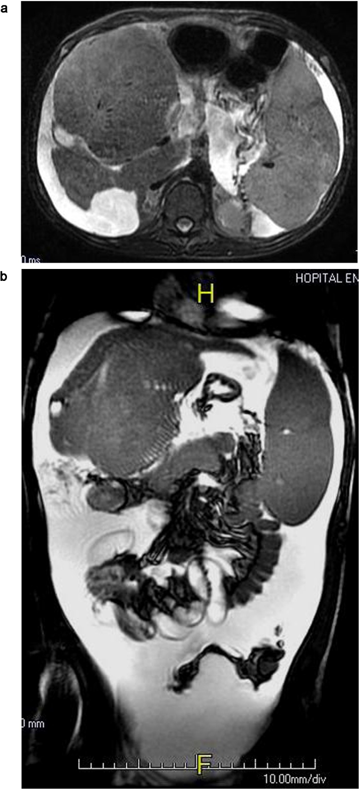
Presentation of case: A 15 year-old girl with Alagille syndrome was referred for liver transplantation. She developed severe cirrhosis with refractory ascites. In the pre-transplant evaluation, imaging studies disclosed liver atrophy with a high density pseudotumor in the segment 4, raising the possibility of a hepatocellular carcinoma. However, behavior of the lesion was highly suggestive of focal compensatory hyperplasia surrounded by an atrophic liver. The patient was registered on the waiting list.
Discussion: Hepatic lesions have been described in Alagille syndrome in isolated case reports, and most of these have been reported to be hepatocellular carcinoma. However, they can be related to an area of focal compensatory hyperplasia in severe cirrhosis. These findings may also explain why progression of liver disease occurs only in 15% of patients.
Conclusion: The presence of a large hepatic nodule Alagille syndrome can be benign in these patients also predisposed to hepatocellular carcinoma. Therefore, cautious evaluation with magnetic resonance imaging study before liver transplantation is mandatory.
Keywords: Alagille syndrome; Case report; Cirrhosis; Focal liver hyperplasia.
Copyright © 2016 The Author(s). Published by Elsevier Ltd.. All rights reserved.
Figures


References
-
- Alagille D., Estrada A., Hadchouel M., Gautier M., Odièvre M., Dommergues J.P. Syndromic paucity of interlobular bile ducts. J. Pediatr. 1987;110:195–200. - PubMed
-
- Emerick K.M., Rand E.B., Goldmuntz E., Krantz I.D., Spinner N.B., Piccoli D.A. Features of Alagille syndrome in 92 patients: frequency and relation to prognosis. Hepatology. 1999;29:822–829. - PubMed
-
- Torizuka T., Tamaki N., Fujita T., Yonekura Y., Uemoto S., Tanaka K. Focal liver hyperplasia in Alagille syndrome: assessment with hepatoreceptor and hepatobiliary imaging. J. Nucl. Med. 1996;37:1365–1367. - PubMed
-
- Adams P.C. Hepatocellular carcinoma associated with arteriohepatic dysplasia. Dig. Dis. Sci. 1986;31:438–442. - PubMed
LinkOut - more resources
Full Text Sources
Other Literature Sources
Research Materials
