. 2016 Sep:102:87-97.
doi: 10.1016/j.biomaterials.2016.06.015. Epub 2016 Jun 7.

Tunable, biodegradable gold nanoparticles as contrast agents for computed tomography and photoacoustic imaging

Affiliations

Tunable, biodegradable gold nanoparticles as contrast agents for computed tomography and photoacoustic imaging

Rabee Cheheltani et al. Biomaterials. 2016 Sep.

Abstract

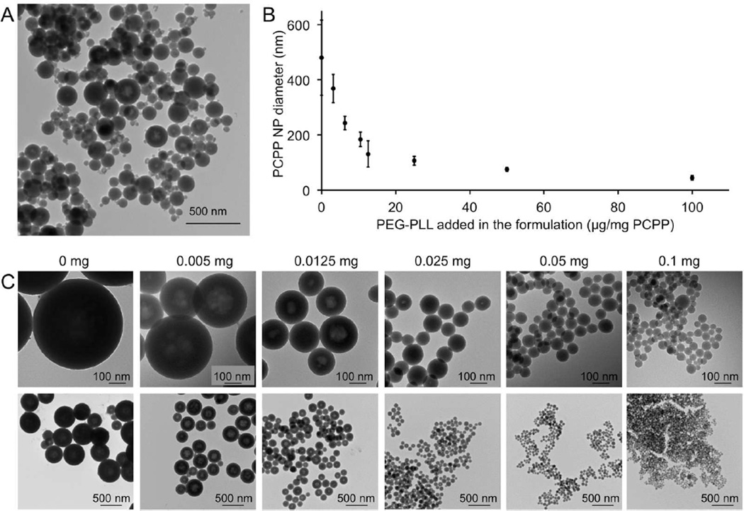
Gold nanoparticles (AuNP) have been proposed for many applications in medicine. Although large AuNP (>5.5 nm) are desirable for their longer blood circulation and accumulation in diseased tissues, small AuNP (<5.5 nm) are required for excretion via the kidneys. We present a novel platform where small, excretable AuNP are encapsulated into biodegradable poly di(carboxylatophenoxy)phosphazene (PCPP) nanospheres. These larger nanoparticles (Au-PCPP) can perform their function as contrast agents, then subsequently break down into harmless byproducts and release the AuNP for swift excretion. Homogeneous Au-PCPP were synthesized using a microfluidic device. The size of the Au-PCPP can be controlled by the amount of polyethylene glycol-polylysine (PEG-PLL) block co-polymer in the formulation. Synthesis of Au-PCPP nanoparticles and encapsulation of AuNP in PCPP were evaluated using transmission electron microscopy and their biocompatibility and biodegradability confirmed in vitro. The Au-PCPP nanoparticles were found to produce strong computed tomography contrast. The UV-Vis absorption peak of Au-PCPP can be tuned into the near infrared region via inclusion of varying amounts of AuNP and controlling the nanoparticle size. In vitro and in vivo experiments demonstrated the potential of Au-PCPP as contrast agents for photoacoustic imaging. Therefore, Au-PCPP nanoparticles have high potency as contrast agents for two imaging modalities, as well as being biocompatible and biodegradable, and thus represent a platform with potential for translation into the clinic.

Keywords: Computed tomography; Contrast agent; Gold nanoparticle; Metal-polymer nanoparticle; Photoacoustic imaging.


Figures

Figure 1
Figure 1
Schematic depiction of biodegradable gold nanoparticles. Small AuNP are incorporated into a biodegradable polyphosphazene (PCPP) resulting in larger nanoparticles with potent contrast agent imaging capabilities. These larger nanoparticles degrade in vivo and release the smaller AuNP for excretion.
Figure 2
Figure 2
A) TEM image of PCPP nanoparticles synthesized without a microfluidic device (bulk synthesis) showing the polydispersity of their sizes. B) Size variation of PCPP nanoparticles synthesized using the microfluidic device as a function of PEG-PLL amount in the formulation. C) TEM images of PCPP nanoparticles synthesized using a microfluidic device with varying amounts of PEG-PLL added in the formulation (presented as mg PEG-PLL per mg of PCPP).
Figure 3
Figure 3
TEM images of Au-PCPP synthesized using microfluidics. A–C) 100 nm Au-PCPP loaded with 0.5 mg (A), 2.5 mg (B) and 5 mg (C) of glutathione coated AuNP per mg PCPP. D–F) TEM images of Au-PCPP loaded with 11-MUA coated AuNP synthesized using 25 µg (D), 12.5 µg (E) and 6.25 µg (F) PEG-PLL per mg PCPP in the formulation.
Figure 4
Figure 4
Selection of a formulation for PA imaging. A) When AuNP are aggregated into Au-PCPP particles, their UV-vis spectra shifts into the NIR region. B) UV-vis measurements of the 11-MUA coated Au-PCPP revealed increasing red shifts into the near infrared by increasing the AuNP loading and size of Au-PCPP. The shift in UV-vis absorption peak is linearly correlated (R2>0.94) with the amount of AuNP loaded into the PCPP nanoparticles. C) TEM image and D) SEM image of the selected formulation i.e. 9 µg PEG-PLL/mg PCPP loaded with 2 mg Au/mg PCPP.
Figure 5
Figure 5
In vitro biocompatibility of Au-PCPP. A) Viability of cells incubated with varying concentrations of Au-PCPP. B) Viability of cells incubated for 8 hours with cell culture media aged with Au-PCPP for different time spans.
Figure 6
Figure 6
A) TEM of Au-PCPP 6 months after synthesis. B) UV/vis spectra of freshly made and one year old Au-PCPP samples. C) SEM and D) TEM of Au-PCPP after 7 days incubation with 10% serum (37 °C). E) Degradation of Au-PCPP in 10% serum (37 °C). F) Effect of Au-PCPP size on degradation. G) TEM of glutathione coated AuNP after release from PCPP. H) TEM of macrophage cells incubated with Au-PCPP for 24 hours showing the release of AuNP inside the cells. I) Release of AuNP from macrophages incubated with Au-PCPP.
Figure 7
Figure 7
CT contrast enhancement with Au-PCPP. A) CT image of Au-PCPP phantoms. B) Linear correlation (R2=0.96) of CT attenuation and Au concentration in Au-PCPP in phantoms. C) Strong CT contrast of Au-PCPP from IM injection in mice.
Figure 8
Figure 8
PA contrast properties of Au-PCPP loaded with 11-MUA coated AuNP. A) Relationship between PA contrast and Au concentration in Au-PCPP and AuNP. B) PA intensity of Au-PCPP compared to AuNP. C) PA contrast arising from Au-PCPP injected IM in mice. The grayscale images on the left are ultrasound images acquired simultaneously with the PA images. The PA images are the red images on the left.

References

    1. Cormode DP, Skajaa T, Fayad ZA, Mulder WJM. Nanotechnology in Medical Imaging: Probe Design and Applications. Arterioscler. Thromb. Vasc. Biol. 2009;29:992–1000. - PMC - PubMed
    1. Kircher MF, Willmann JK. Molecular Body Imaging: MR Imaging, CT, and US. Part I. Principles. Radiology. 2012;263:633–643. - PMC - PubMed
    1. Jokerst JV, Lobovkina T, Zare RN, Gambhir SS. Nanoparticle PEGylation for Imaging and Therapy. Nanomedicine (Lond) 2011;6:715–728. - PMC - PubMed
    1. Thaxton CS, Rink JS, Naha PC, Cormode DP. Lipoproteins and Lipoprotein Mimetics for Imaging and Drug Delivery. Adv. Drug Deliv. Rev. 2016 - PMC - PubMed
    1. Shapiro EM. Biodegradable, Polymer Encapsulated, Metal Oxide Particles for MRI-Based Cell Tracking. Magn. Reson. Med. 2015;73:376–389. - PMC - PubMed

Publication types