. 2016 Jun 21:6:28297.
doi: 10.1038/srep28297.

Zebrafish cyclin Dx is required for development of motor neuron progenitors, and its expression is regulated by hypoxia-inducible factor 2α

Affiliations

Zebrafish cyclin Dx is required for development of motor neuron progenitors, and its expression is regulated by hypoxia-inducible factor 2α

Huang-Wei Lien et al. Sci Rep. .

Abstract

Cyclins play a central role in cell-cycle regulation; in mammals, the D family of cyclins consists of cyclin D1, D2, and D3. In Xenopus, only homologs of cyclins D1 and D2 have been reported, while a novel cyclin, cyclin Dx (ccndx), was found to be required for the maintenance of motor neuron progenitors during embryogenesis. It remains unknown whether zebrafish possess cyclin D3 or cyclin Dx. In this study, we identified a zebrafish ccndx gene encoding a protein which can form a complex with Cdk4. Through whole-mount in situ hybridization, we observed that zccndx mRNA is expressed in the motor neurons of hindbrain and spinal cord during development. Analysis of a 4-kb promoter sequence of the zccndx gene revealed the presence of HRE sites, which can be regulated by HIF2α. Morpholino knockdown of zebrafish Hif2α and cyclin Dx resulted in the abolishment of isl1 and oligo2 expression in the precursors of motor neurons, and also disrupted axon growth. Overexpression of cyclin Dx mRNA in Hif2α morphants partially rescued zccndx expression. Taken together, our data indicate that zebrafish cyclin Dx plays a role in maintaining the precursors of motor neurons.


Figures
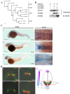
Figure 1
Figure 1. Zebrafish cyclin Dx is a member of the cyclin D family, and zccndx is expressed during early development.
(A) Phylogenetic tree analysis of zccndx with other cyclin D proteins. Four groups of the cyclin D family, ccnD1, D2, D3, and Dx, exhibited perfect separation. The zccndx protein was grouped with Xenous ccndx. Analysis was performed using the parsimony method with 1000 bootstraps. (B) The zccndx protein binds to Cdk4. COS-1 cells were cotransfected with the indicated constructs, and cell lysates were co-immunoprecipitated (IP) with anti-HA monoclonal antibody; the anti-Myc monoclonal Ab was used for detection. The immunoblot was probed using the indicated antibodies. (C) At 24 hpf, zccndx mRNA was observed to be expressed in the motor neurons of hindbrain and spinal cord (a and a′). At 48 hpf, zccndx mRNA was also expressed in the hindbrain, but not in the spinal cord (b and b′). At 72 hpf, zccndx mRNA was weakly expressed in the hindbrain, with no signal in the spinal cord (c and c′). Dorsal views of zccndx mRNA signal in the hindbrain are shown (a′–c′). Scale bar, 100 mm. (D) Sections were made in the hindbrain at 22 hpf. The zccndx signal is shown in green and the isl1 (panel a), olig2 (panel b), and vsx1 (panel c), and nkx6.1 (panel d) mRNA signals are shown in red. The cartoon (panel e) shows the location of zccndx (green), isl1 (purple), olig2 (orange), vsx1 (blue), and nkx6.1 (red) signals. The Shh (blue) signaling gradient has been created from the floor plate to diffuse along the ventral tube while BMP/Wnt (purple) gradient shows opposite direction. The interaction of the dorsalizing and ventralizing signals defines the proper position of motoneurons. Scale bar, 100 mm.
Figure 2
Figure 2. Morpholino (MO) knockdown of zccndx affects motor neuron development.
(A) MO knockdown of zccndx reduced the olig2 and isl1 signals in motor neurons (panels a, b, e, and f); rescue experiments performed by co-injecting ccndx mRNA are shown in panels c and g. Panels d and h: graphs showing the qPCR analysis confirmed the results of morphants with signals lower than control MO-injected embryos at 24 hpf. **P < 0.05. Scale bar, 100 mm. (B) The ccndx morphant showed a significantly higher ratio of embryos with lower numbers of GFP-labeled primary motoneurons than the control MO-injected embryos (panels a and b) in the Tg:(isl1:GFP) transgenic line. The ccndx mRNA recue experiment is shown in panel c. Panel d: the Statistical charts represent the quantitative results of GFP-labeled primary motoneurons cells at 24 hpf. (C) Labeling of primary motor axons with Znp1 antibody revealed that CaP axon branches were shorter in ccndx morphants than in control morphants (panels a and b); the rescue experiment is shown in panel c. Panel d: graphs showing the percentages of morphants with primary motor defects as compared to control MO-injected embryos at 24 hpf in panels d and h. *P < 0.05.
Figure 3
Figure 3. The role of zccndx in cell proliferation and regulation of the numbers of motor neuron progenitors (pMNs).
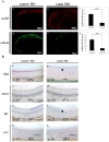
(A) Confocal images showing lateral views of the heads of control-MO-injected (panel a) or ccndx-MO-injected (panel b) embryos stained with anti-pH3 (red in panels a and b) or anti-BrdU (green in d and e). Panels c and f: graphs showing the number of proliferating cells (pH3 or BrdU positive) at 24 hpf. The asterisks indicate significant differences between the control and experimental morphants. The n value is indicated. (B) Whole–mount in situ hybridization of motor neurons and interneuron markers in control-MO and zccndx ATG-MO-injected embryos. Probes against olig2 (a and b), nkx6.1(c and d), isl1 (e and f), and vsx1 (g and h) were used for detection. control MO-injected embryos are shown in panels a, c, e and g while ccndx ATG-MO-injected embryos are shown in panels b, d, f and h. Motor neurons (MN) were lost (panels b, f, lost MNs indicated by arrowheads), while interneurons (IN) (panels d and h) were unaffected. Scale bar, 100 mm.
Figure 4
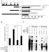
Figure 4. Expression of the ccndx gene is regulated by the HIF2α transcription factor.
(A) The promoter region of the ccndx gene contains several HRE sites, as indicated in the schematic diagram (top left). The 2.5-kb, 1-kb, and 0.7-kb promoters are all activated by HIF2α and arnt1a transcription factors. (B) The HIF2α and ARNT1α proteins co-activate ccndx gene expression through two HRE sites in the 700bp promoter region. Data represent mean ± s.d. (n = 3). (C) The DNA binding ability of HIF2α and the ARNT1α complex were confirmed by EMSA. Nuclear extract (NE) from pcDNA3-HA (Con) or pcDNA3-Hif2α-HA and pcDNA3-Arnt1α-HA (HIF2α + ARNT1α) transfection were indicated. Two HRE oligonucleotides (as indicated) were used as probes and normal (Cold) or mutant (Mut) oligonucleotides were used for competition. The asterisks in lanes 2, 4, 6, and 8 indicate the specific binding complex.
Figure 5
Figure 5. The HIF2α transcription factor helps regulate ccndx gene expression and subsequent motor neuron development.
(A) MO knockdown of hif2a reduced the olig2 and isl1 signals in motor neurons (panels a, b, e, and f); co-injection of ccndx mRNA partially rescued this phenotype (panels c and g). Panels d and h: graphs showing the qPCR analysis confirmed the results of morphants with signals lower than control MO-injected embryos at 24 hpf. *P < 0.05. Scale bar, 100 mm. (B) A significantly higher proportion of hif2a morphant embryos had lower numbers of GFP-labeled primary motoneurons as compared to control MO-injected embryos (panels a and b) in the Tg:(isl1:GFP) transgenic line. The effect of rescue experiments with ccndx mRNA is shown in panel c. Quantitative data at 24 hpf are shown in panel d. (C) A significantly higher proportion of hif2a morphant embryos exhibited shorter CaP axons branches as compared to control MO-injected embryos (panels a and b). The effect of rescue experiments with ccndx mRNA is shown in panel c. Quantitative data at 24 hpf are shown in panel d. *P < 0.05.
Figure 6
Figure 6. Mouse cyclin D1, D2, and D3 cannot compensate for the loss of zebrafish cyclin Dx.
(A) MO knockdown of ccndx reduced the isl1 and olig2 signals in motor neurons (panels a, b, g, and h); co-injection of mouse ccnd1, ccnd2, and ccnd3 mRNA had no effect, as shown in panels c to e and i to k. Panels r and l: graphs showing the qPCR analysis confirmed the results of morphants with signals lower than control MO-injected embryos at 24 hpf. *P < 0.05. Scale bar, 100 mm. (B) A significant number of ccndx morphant embryos contained fewer GFP-labeled primary motoneurons as compared to ccndx morphants (panels a and b) in the Tg:(isl1:GFP) transgenic line, and co-injection of mouse ccnd1, ccnd2, or ccnd3 mRNA was unable to rescue this defect (panels c, d, and e). Panel f: graph showing the quantitative results of GFP-labeled primary motoneurons cells at 24 hpf. (C) A significant number of ccndx morphant embryos exhibited shorter primary motor axons as compared to ccndx morphants (panels a and b), and co-injection of mouse ccnd1, ccnd2, or ccnd3 mRNA was unable to rescue this defect (panels c, d, and e). Panel f: graph showing the proportion of embryos with signals lower than those in control MO-injected embryos at 24 hpf.

References

    1. Hayles J. & Nurse P. Cell cycle regulation in yeast. J Cell Sci Suppl 4, 155–170 (1986). - PubMed
    1. Lee M. & Nurse P. Cell cycle control genes in fission yeast and mammalian cells. Trends Genet 4, 287–290(1988). - PubMed
    1. Pelech S. L., Sanghera J. S. & Daya-Makin M. Protein kinase cascades in meiotic and mitotic cell cycle control. Biochem Cell Biol 68, 1297–1330 (1990). - PubMed
    1. Maller J. L. Xenopus oocytes and the biochemistry of cell division. Biochemistry 29, 3157–3166 (1990). - PubMed
    1. Norbury C. & Nurse P. Animal cell cycles and their control. Annu Rev Biochem 61, 441–470 (1992). - PubMed

Substances