. 2016 Jun 21:6:28126.
doi: 10.1038/srep28126.

The use of platelet-rich fibrin combined with periodontal ligament and jaw bone mesenchymal stem cell sheets for periodontal tissue engineering

Affiliations

The use of platelet-rich fibrin combined with periodontal ligament and jaw bone mesenchymal stem cell sheets for periodontal tissue engineering

Zhong-Shan Wang et al. Sci Rep. .

Abstract

Periodontal regeneration involves the restoration of at least three unique tissues: cementum, periodontal ligament tissue (PDL) and alveolar bone tissue. Here, we first isolated human PDL stem cells (PDLSCs) and jaw bone mesenchymal stem cells (JBMSCs). These cells were then induced to form cell sheets using an ascorbic acid-rich approach, and the cell sheet properties, including morphology, thickness and gene expression profile, were compared. Platelet-rich fibrin (PRF) derived from human venous blood was then fabricated into bioabsorbable fibrin scaffolds containing various growth factors. Finally, the in vivo potential of a cell-material construct based on PDLSC sheets, PRF scaffolds and JBMSC sheets to form periodontal tissue was assessed in a nude mouse model. In this model, PDLSC sheet/PRF/JBMSC sheet composites were placed in a simulated periodontal space comprising human treated dentin matrix (TDM) and hydroxyapatite (HA)/tricalcium phosphate (TCP) frameworks. Eight weeks after implantation, the PDLSC sheets tended to develop into PDL-like tissues, while the JBMSC sheets tended to produce predominantly bone-like tissues. In addition, the PDLSC sheet/PRF/JBMSC sheet composites generated periodontal tissue-like structures containing PDL- and bone-like tissues. Further improvements in this cell transplantation design may have the potential to provide an effective approach for future periodontal tissue regeneration.


Conflict of interest statement

The authors declare no competing financial interests.

Figures

Figure 1
Figure 1. The differences in proliferative capacity and immunophenotype between human periodontal ligament (PDL) stem cells (PDLSCs) and human jaw bone mesenchymal stem cells (JBMSCs).
(A) The initial PDLSCs and JBMSCs were cultured in six-well dishes containing complete α-MEM. (B) Representative images of crystal violet staining of the colonies of PDLSCs and JBMSCs and a comparison of the number of colonies formed by PDLSCs and JBMSCs (*P < 0.05 indicates a significant difference compared with PDLSCs). (C) Growth curves for PDLSCs and JBMSCs, as determined by a cell counting kit-8 (CCK-8) assay (*P < 0.05 and **P < 0.01 indicate significant differences compared with PDLSCs at the same time point). (D) Flow cytometric analysis indicated that the PDLSCs and JBMSCs were positive for mesenchymal-associated markers CD29, CD146 and STRO-1 but negative for hematopoietic markers CD34 and CD45 (*P < 0.05 indicates a significant difference compared with JBMSCs).
Figure 2
Figure 2. The differences in multi-directional differentiation potential between PDLSCs and JBMSCs.
(A) Representative images showing the differences in multipotent differentiation between the two cell types. Lipid vacuoles of different sizes and numbers were observed after 2 weeks of adipogenic induction (Oil Red O staining, scale bar = 200 μm). Data analysis was performed on the relative level of Oil Red O dye absorption between PDLSCs and JBMSCs (*P < 0.05 indicates a significant difference compared with PDLSCs). (B) Mineralized nodules of different sizes and numbers were formed after 4 weeks of osteogenic induction (Alizarin Red S staining, scale bar = 100 μm). Data analysis was performed on the relative level of Alizarin Red S dye absorption between PDLSCs and JBMSCs (*P < 0.05 indicates a significant difference compared with PDLSCs). (C) The difference in osteogenic differentiation capacity between PDLSCs and JBMSCs in vivo at eight weeks post-implantation. The three-dimensional (3-D) reconstruction images of grafts were obtained via micro-CT, and the results indicated that the JBMSC/HA/TCP group (more red areas) displayed a higher density than did the PDLSC/HA/TCP group (more green areas). The equivalent bone density was quantitatively analyzed through statistical analysis (*P < 0.05 indicates a significant difference compared with the PDLSC/HA/TCP group). (D) Hematoxylin and eosin (H&E) staining of the sections after decalcification showed that the PDLSC/HA/TCP group produced predominantly PDL-like structures (green arrow) and some cementum-like structures (red arrows). By contrast, the JBMSC/HA/TCP group displayed predominantly bone-like structures that varied in appearance and size (blue arrows). The mixed HA/TCP clumps are indicated by yellow arrows in both groups. (E) The differences in gene expression between PDLSCs (P2) and JBMSCs (P2) (*P < 0.05 and **P < 0.01 indicate significant differences compared with PDLSCs).
Figure 3
Figure 3. Characterization of the PDLSC and JBMSC sheets.
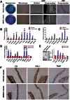
(A) Representative images of PDLSC and JBMSC sheets formed in culture dishes (general view), as observed under an inverted microscope (cell arrangement, scale bar = 100 μm), scanning electron microscopy (SEM; cell arrangement and connections), and H&E staining (cross-section, scale bar = 10 μm). (B) The relative mRNA expression profiles of PDLSC sheets compared to PDLSCs (*P < 0.05 and **P < 0.01 indicate significant differences compared with PDLSCs). (C) The relative mRNA expression profiles of JBMSC sheets compared to JBMSCs (*P < 0.05 and **P < 0.01 indicate significant differences compared with JBMSCs). (D) The relative mRNA expression profiles of JBMSC sheets compared to PDLSC sheets (*P < 0.05 and **P < 0.01 indicate significant differences compared with PDLSC sheets). (E) The expression levels of periodontal tissue-specific proteins (COL I and COL III), calcification-related proteins (OPN and OCN), and cementum tissue-related proteins (CEMP1 and CAP) in PDLSC and JBMSC sheets as determined by quantitative western blot analysis (*P < 0.05 and **P < 0.01 indicate significant differences compared with PDLSC sheets). (F) Immunohistological characteristics of PDLSC and JBMSC sheets. Both types of cell sheets displayed strong positive immunohistochemical staining using anti-human FN1 and anti-human COL I antibodies. Both sheets also displayed weakly positive staining for TNAP.
Figure 4
Figure 4. Characterization of transplanted biomaterials (platelet-rich fibrin (PRF), human treated dentin matrix (TDM) and HA/TCP frameworks) and their combined application for periodontal regeneration in a nude mouse model.
(A) (a,b) PRF clot after centrifugation. (c,d) The ultrastructure of the PRF as observed by SEM. In the top region (c) of the PRF, the 3-D network predominantly comprised fibrin fibers (red arrow) and a few fibrillae with smaller diameters (blue arrow), and few platelets or cells were detected. In the lower region of the PRF (d), numerous platelets (red arrow) and leukocytes (blue arrow) and some red blood cells (yellow arrow) were embedded in the network. (B) The macroscopic and microscopic appearance of the human TDM and HA/TCP scaffolds. (a,b) The macroscopic appearance of a TDM and an HA/TCP scaffold with a length of 1.0 cm. TDM was fabricated using a previously described method to simulate dentin, and HA/TCP frameworks with a tooth root-like shape were used to simulate alveolar bone. (c) The appearance of the HA/TCP frameworks of varying size as observed by SEM. The HA/TCP particles were loosely and irregularly arranged, and there were many pores between the particles. (d) The appearance of TDM as dentinal tubules arranged in an orderly manner as observed by SEM. (C) (a) Cell sheets were detached after maturation and wrapped in eight layers on the surfaces of the TDM (b). A ring-shaped interspace was formed after insertion of the TDM into the HA/TCP framework (c), and this void provided space for transplanted cell sheets and PRF (d).
Figure 5
Figure 5. Mimicking periodontal regeneration in a nude mouse model using a PRF scaffold combined with PDLSC and JBMSC sheets.
Representative images of periodontal tissue regeneration in the different groups. In group 1 (PDLSC sheets/PRF/PDLSC sheets), abundant collagen fibers (PDL) were produced, and blood vessels (yellow arrows) crossing through the tissue were present, but there was no evidence of bone-like tissue formation. In group 2 (JBMSC sheets/PRF/JBMSC sheets), abundant bone-like tissues (B) as well as some newly formed vessels (yellow arrows) were observed; however, fewer collagen fibers were present in group 2 than in group 1. In group 3 (PDLSC sheets/PRF/JBMSC sheets), both PDL- and bone-like structures were produced. Briefly, a dense layer of connective tissue covered the TDM surface. Outside the PDL-like tissue, several layers of bone-like tissue (B) were observed (scale bar = 100 μm).

References

    1. Albandar J. M. Aggressive and Acute Periodontal Diseases. Periodontol. 65, 7–12 (2014). - PubMed
    1. Su F. et al.. Enhancement of periodontal tissue regeneration by transplantation of osteoprotegerin-engineered periodontal ligament stem cells. Stem Cell Res. Ther. 6, 1–14 (2015). - PMC - PubMed
    1. Manjunath B. C., Praveen K., Chandrashekar B. R., Rani R. M. & Bhalla A. Periodontal infections: a risk factor for various systemic diseases. Natl. Med. J. India. 24, 214–219 (2011). - PubMed
    1. Chen F. M. et al.. A review on endogenous regenerative technology in periodontal regenerative medicine. Biomaterial 31, 7892–7927 (2010). - PubMed
    1. Bosshardt D. D. & Sculean A. Does periodontal tissue regeneration really work? Periodontol. 51, 208–219 (2009). - PubMed

Publication types

MeSH terms

LinkOut - more resources