Review
. 2016 Sep 15;594(18):5055-77.
doi: 10.1113/JP270192. Epub 2016 Jul 21.

Arteriolar oxygen reactivity: where is the sensor and what is the mechanism of action?

Affiliations
Review

Arteriolar oxygen reactivity: where is the sensor and what is the mechanism of action?

William F Jackson. J Physiol. .

Abstract

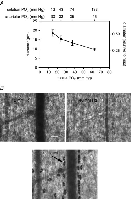
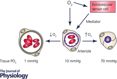
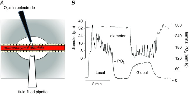
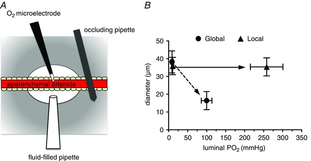
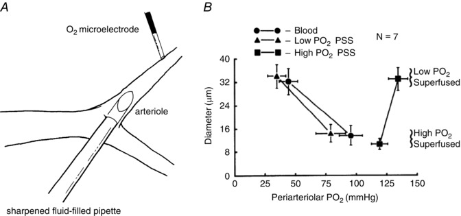
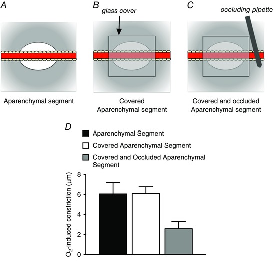
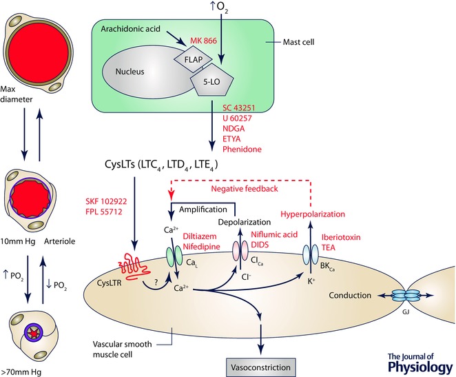
Arterioles in the peripheral microcirculation are exquisitely sensitive to changes in PO2 in their environment: increases in PO2 cause vasoconstriction while decreases in PO2 result in vasodilatation. However, the cell type that senses O2 (the O2 sensor) and the signalling pathway that couples changes in PO2 to changes in arteriolar tone (the mechanism of action) remain unclear. Many (but not all) ex vivo studies of isolated cannulated resistance arteries and large, first-order arterioles support the hypothesis that these vessels are intrinsically sensitive to PO2 with the smooth muscle, endothelial cells, or red blood cells serving as the O2 sensor. However, in situ studies testing these hypotheses in downstream arterioles have failed to find evidence of intrinsic O2 sensitivity, and instead have supported the idea that extravascular cells sense O2 . Similarly, ex vivo studies of isolated, cannulated resistance arteries and large first-order arterioles support the hypotheses that O2 -dependent inhibition of production of vasodilator cyclooxygenase products or O2 -dependent destruction of nitric oxide mediates O2 reactivity of these upstream vessels. In contrast, most in vivo studies of downstream arterioles have disproved these hypotheses and instead have provided evidence supporting the idea that O2 -dependent production of vasoconstrictors mediates arteriolar O2 reactivity, with significant regional heterogeneity in the specific vasoconstrictor involved. Oxygen-induced vasoconstriction may serve as a protective mechanism to reduce the oxidative burden to which a tissue is exposed, a process that is superimposed on top of the local mechanisms which regulate tissue blood flow to meet a tissue's metabolic demand.

Keywords: arterioles; microcirculation; oxygen; oxygen sensing; vasoconstriction; vasodilatation.


Figures

Figure 1
Figure 1. Arteriolar O2 reactivity in superfused microvascular preparations
A, data from Klitzman et al. (1982). Data are mean ± SEM diameters of arterioles in hamster cremaster muscles when exposed to solutions equilibrated with gases containing different PO2values and 5% CO2 (solution PO2 values, upper x‐axis labels). The PO2 values shown were measured with O2 microelectrodes in the free solution flowing over the preparations. The tissue PO2 values shown on the lower x‐axis were measured at the midpoint between the venous ends of two capillaries. The arteriolar PO2 values (top x‐axis labels) were estimated from haemoglobin oxygen saturation measurements in hamster cheek pouch (Jackson & Duling, 1983). B, typical O2‐induced vasoconstriction in a hamster cheek pouch preparation exposed to superfusion solutions with approximate PO2values as indicated using methods as described (Jackson, 1987). C, same vessel as in B after exposure to ruthenium red (0.001%) to label mast cells (Shepherd & Duling, 1995). Scale bar in B, 25 μm.
Figure 2
Figure 2. Schematic diagram of O2 signalling in the microcirculation
Oxygen in the environment of arterioles can act directly on the arteriolar wall or on cells in the lumen to produce a vasomotor effect (dilatation in the case of reduced PO2, or constriction in the case of elevated PO2). Alternatively, changes in PO2 may be sensed by extravascular cells (parenchymal cells, mast cells, nerves, etc.), a mediator produced, which then acts on the vessel wall to produce the appropriate vasomotor effect. The PO2 values shown below the cross section of the arterioles refer to tissue PO2 in a superfused, intravital preparation measured at the midpoint between the venous ends of two capillaries as reference values, only.
Figure 3
Figure 3. Local increases in PO2 have no effect on arteriolar tone
Data shown are modified from Jackson (1987). A, schematic diagram of a segment (1–8 mm) of an arteriole in a hamster cheek pouch from which the parenchyma has been surgically removed (aparenchymal arteriole) to obviate effects of local PO2 changes on parenchymal and other extravascular cells. A Whalen‐type recessed tip O2 microelectrode was inserted through the wall of the vessel into the lumen as shown to monitor luminal PO2. A temperature‐controlled micropipette filled with PSS equilibrated with varied PO2 (fluid‐filled micropipette) was positioned opposite the O2 microelectrode. Pressurization of the fluid‐filled micropipette ejected the O2‐equilibrated solution onto the surface of the arteriole to produce a local change in PO2 . The entire cheek pouch preparation was superfused with PSS, the PO2 of which could be varied to produce global changes in PO2 that affected both the aparenchymal arteriole and all other vessels and cell types in the cheek pouch. The diameter of the arteriole was measured by intravital video microscopy. B, results from a typical experiment in which either local increases in PO2 were produced using the fluid‐filled pipette (Local) or global increases in PO2 were produced by changing the PO2 of the entire superfusate (Global) (replotted data are from Fig. 3 in Jackson, 1987). Local increases in PO2 that effectively changed the PO2 across the wall of the arteriole had no significant effect on arteriolar diameter, whereas a global increase in PO2 produced sustained vasoconstriction. These data suggest that components of the arteriolar wall (endothelial cells, smooth muscle cells or perivascular nerves) are not the sensor cells responsible for arteriolar O2 reactivity in the hamster cheek pouch. See Jackson (1987) for details.
Figure 4
Figure 4. Local increases in PO2 have no effect on arteriolar tone in occluded arterioles
A, a preparation similar to that depicted in Fig. 3, but with the inclusion of an occluding pipette that was pressed down on the arteriole to eliminate blood flow through the aparenchymal segment. B, summary data from these experiments (data are means ± SEM, n = 5). Local changes in PO2 across the arteriolar wall produced by the fluid‐filled pipette (Local) were ineffective in producing arteriolar constriction. However, raising the PO2 of the superfusate over the entire preparation (Global) produced consistent, sustained arteriolar constriction. These data, along with those shown in Fig. 3, suggest that components of the arteriolar wall (endothelial cells, smooth muscle cells or perivascular nerves) are not the sensor cells responsible for arteriolar O2 reactivity in the hamster cheek pouch. Data are replotted from Fig. 4 B in Jackson (1987); see this reference for more details.
Figure 5
Figure 5. Perfusion of arterioles in situ with solutions equilibrated with high PO2 has no effect on arteriolar tone
A and B are reproduced from Jackson (1987). A, schematic diagram of a hamster cheek pouch arteriole in which a sharpened fluid‐filled pipette has been inserted through the wall of an arteriole allowing perfusion of the arteriole with PSS equilibrated with varied PO2. The entire cheek pouch preparation was superfused with PSS to allow global changes in PO2. B, summary data (means ± SEM). Perfusion of the arterioles with solutions equilibrated with high or low PO2 had no significant effect on arteriolar diameter. Only when the global PO2 was elevated via the superfusate did the arterioles constrict (compare low PO2 superfusate points with high PO2 superfusate points). These data suggest that components of the arteriolar wall (endothelial cells, smooth muscle cells or perivascular nerves) are not the sensor cells responsible for arteriolar O2 reactivity in the hamster cheek pouch. See Jackson (1987) for details.
Figure 6
Figure 6. Covering aparenchymal segments with glass and occluding them to eliminate blood flow does not eliminate arteriolar O2 reactivity
A, schematic diagram of an aparenchymal segment in which the parenchyma has been removed from a long segment of hamster cheek pouch arteriole as described by Jackson & Duling (1983). Elevation of the PO2 of the solution flowing over the preparation from 12 to 150 mmHg resulted in arteriolar constriction as depicted in D. As shown in B, subsequent covering of the aparenchymal segment with a piece of glass coverslip (sealed in place with silicone grease), to eliminate contact of the arteriole with the superfusate, had no effect on O2‐induced constriction as shown in D. To eliminate blood flow through the covered aparenchymal segments, an occluding pipette was used as shown in C. Despite the lack of access to the superfusate and flowing blood, these covered and occluded aparenchymal segments retained significant O2 reactivity as shown in D. These data suggest that the constriction induced by elevated PO2 can be conducted along the arteriolar wall. See Jackson & Duling (1983) for details.
Figure 7
Figure 7. The site and mechanism of action of O2 in the hamster cheek pouch
Schematic diagram depicting a mast cell (the proposed sensor site in the cheek pouch), a smooth muscle cell replete with receptors for cysteinyl leukotrienes (CysLTRs) and ion channels involved in arteriolar O2 reactivity in the cheek pouch. Elevated PO2 is sensed by the 5‐lipoxygenase (5‐LO) in the nuclear membrane of periarteriolar mast cells that decorate arterioles in this tissue (see Fig. 1). This results in conversion of arachidonic acid to cysteinyl leukotrienes (CysLTs) such as LTC4, LTD4 and LTE4 through a process that involves presentation of the arachidonic acid to the 5‐LO by the 5‐LO‐activating protein (FLAP). This process can be inhibited by drugs such as MK 866, that blocks interaction of FLAP with the 5‐LO, or SC 43251, U 60257, nordihydroguaiaretic acid (NDGA), eicosatetraynoic acid (ETYA) or phenidone, inhibitors of the 5‐LO. The CysLTs then bind to and activate CysLTRs on vascular smooth muscle cells to induce vasoconstriction. CysLTR antagonists such as SKF 102922 or FPL 55712 can inhibit this step in the process. Activation of CysLTRs results in activation of L‐type Ca2+ channels (CaL), Ca2+ influx, an increase in intracellular Ca2+ and vasoconstriction, which can be antagonized by CaL blockers such as diltiazem or nifedipine. The increase in Ca2+ activates Ca2+‐activated Cl channels (ClCa). The resulting efflux of Cl through these channels causes membrane depolarization, further activating CaL and amplifying the initial signal. Blockers of ClCa channels such as niflumic acid or DIDs can inhibit this step in the process. The increase in intracellular Ca2+ and the membrane depolarization due to activation of CaL and ClCa activates large conductance, Ca2+‐activated K+ channels (BKCa). The efflux of K+ through BKCa channels blunts the depolarizing effects of activation of CaL and ClCa providing a degree of negative feedback, and limiting membrane depolarization. This step in the process can be inhibited by iberiotoxin or tetraethylammonium ions (TEA). Oxygen‐induced smooth muscle depolarization can be conducted along the vessel wall by gap junctions (GJ) supporting the conduction of O2‐induced vasoconstriction that has been observed experimentally.
Figure 8
Figure 8. The site and mechanism of action of O2 in cremaster muscle
Schematic diagram of a striated muscle fibre (the proposed sensor cell in this tissue) and a smooth muscle cell replete with ion channels that may be involved in arteriolar O2 reactivity in this tissue. Elevated PO2 is sensed by cytochrome P4504A/4F ω‐hydroxylase (CYP4A/4F) located in the endoplasmic reticulum of striated muscle fibres, resulting in conversion of arachidonic acid into 20‐HETE, a process that can be inhibited by 17‐ODYA or DDMS. 20‐HETE then acts on smooth muscle cells to induce Ca2+ influx through L‐type Ca2+ channels (CaL), an increase in intracellular Ca2+ and vasoconstriction. As indicated by the ‘?’ next to the arrow connecting 20‐HETE and the smooth muscle cell, the precise receptor for 20‐HETE that is responsible for O2 reactivity is unclear because 20‐HETE has been proposed to close large conductance, Ca2+‐activated K+ channels (BKCa), which would lead to membrane depolarization activating CaL. In contrast, other studies suggest that BKCa serve a negative feedback role as they do in the cheek pouch. As in the cheek pouch it is proposed that O2‐induced depolarization of smooth muscle cells can be conducted along the vessel wall through gap junctions (GJ), consistent with the observed conducted vasoconstriction that has been observed experimentally. 6(Z),15(Z)‐20‐HEDE, 20‐hydroxy‐6Z,15Z‐eicosadienoic acid.

Similar articles

Cited by

References

    1. Aalkjaer C & Lombard JH (1995). Effect of hypoxia on force, intracellular pH and Ca2+ concentration in rat cerebral and mesenteric small arteries. J Physiol 482, 409–419. - PMC - PubMed
    1. Abu‐Soud HM, Ichimori K, Presta A & Stuehr DJ (2000). Electron transfer, oxygen binding, and nitric oxide feedback inhibition in endothelial nitric‐oxide synthase. J Biol Chem 275, 17349–17357. - PubMed
    1. Baez S (1973). An open cremaster muscle preparation for the study of blood vessels by in vivo microscopy. Microvasc Res 5, 384–394. - PubMed
    1. Baron JF, Vicaut E, Hou X & Duvelleroy M (1990). Independent role of arterial O2 tension in local control of coronary blood flow. Am J Physiol 258, H1388–H1394. - PubMed
    1. Bartlett IS & Segal SS (2000). Resolution of smooth muscle and endothelial pathways for conduction along hamster cheek pouch arterioles. Am J Physiol Heart Circ Physiol 278, H604–H612. - PubMed

Publication types