

Oesophageal and gastric varices: historical aspects, classification and grading: everything in one place
- PMID: 27324725
- PMCID: PMC4976684
- DOI: 10.1093/gastro/gow018
Oesophageal and gastric varices: historical aspects, classification and grading: everything in one place
Abstract
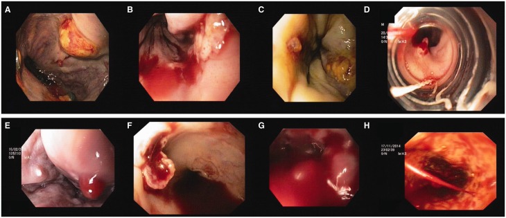
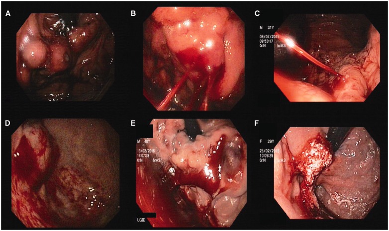
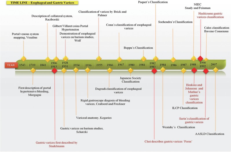
Variceal disease and its management are of the utmost importance in the treatment of portal hypertension. Current guidelines are universal for management of variceal disease in portal hypertension. Classification and grading systems are numerous and differ according to geographical location. In this exhaustive review, the historical aspects of variceal disease, its classification and the grading systems in use are discussed, with self-explanatory tables and timelines. A better and clear understanding of the evolution of portal hypertension and variceal disease is provided.
Keywords: classification; gastric varices; oesophageal varices; portal hypertension.
© The Author(s) 2016. Published by Oxford University Press and Sixth Affiliated Hospital of Sun Yat-Sen University.
Figures


References
-
- Eck NV, Voen Med J. Vol. 130. 1877. K voprosu o perevyazkievorot noisveni: Predvaritelnoyeso obschjenye; pp. 1–2. (Ligature of the portal vein, trans. Child C.G. III: Surg Gynecol Obstet 1953;96:375–76.
-
- Vesalius A. De Humani Corporis Fabrica Basileae. 1543
-
- Morgagni JB. The seats and causes of diseases. New York: Hafner Publishing, 1719, 56–57.
-
- Raciborski A. Histoire de decouvertes relatives au systeme veineux, envisage dans le rapport anatomique, physiologique, pathologique et therapeutique depuis Morgagni jusqu’ànos jours. Mem Acad R Med 1841.
-
- Sappey P. Memoire surun point d’anatomie pathologique relatifal’ histoire de la cirrhose. MemAcad Imp Med 1859.
Publication types
LinkOut - more resources
Full Text Sources
Other Literature Sources
