Review
. 2016 Oct;139(Pt 10):2590-2599.
doi: 10.1093/brain/aww141. Epub 2016 Jun 20.

Cervical dystonia: a neural integrator disorder

Affiliations
Review

Cervical dystonia: a neural integrator disorder

Aasef G Shaikh et al. Brain. 2016 Oct.

Abstract

Ocular motor neural integrators ensure that eyes are held steady in straight-ahead and eccentric positions of gaze. Abnormal function of the ocular motor neural integrator leads to centripetal drifts of the eyes with consequent gaze-evoked nystagmus. In 2002 a neural integrator, analogous to that in the ocular motor system, was proposed for the control of head movements. Recently, a counterpart of gaze-evoked eye nystagmus was identified for head movements; in which the head could not be held steady in eccentric positions on the trunk. These findings lead to a novel pathophysiological explanation in cervical dystonia, which proposed that the abnormalities of head movements stem from a malfunctioning head neural integrator, either intrinsically or as a result of impaired cerebellar, basal ganglia, or peripheral feedback. Here we briefly recapitulate the history of the neural integrator for eye movements, then further develop the idea of a neural integrator for head movements, and finally discuss its putative role in cervical dystonia. We hypothesize that changing the activity in an impaired head neural integrator, by modulating feedback, could treat dystonia.

Keywords: cerebellum; integrator; midbrain; nystagmus; tremor.


Figures

None
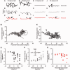
Dysfunction of the oculomotor neural integrator leads to gaze-induced nystagmus. Shaikh et al. briefly recapitulate the basic principles behind neural integration, before developing the idea of an analogous neural integrator for head movements and the putative role of such an integrator in cervical dystonia.
Figure 1
Figure 1
Gaze-evoked nystagmus of the eyes. ( AC ) Example of gaze-evoked nystagmus measured during a 5-s epoch as the subject with a cerebellar lesion attempted to fixate gaze to the right ( A ), straight-ahead ( B ), or to the left ( C ). The nystagmus is characterized by slow phases and rapid corrective quick phases. In a slow phase the eyes drift toward the null ( B ). Slow-phase velocity systematically changes with eye-in-orbit orientation ( D ). Each circle in D depicts one cycle of gaze-evoked nystagmus with the slow-phase velocity on the y -axis while the corresponding eye-in-orbit position is on the x -axis. Positive signs depict the rightward direction, while negative is leftward.
Figure 2
Figure 2
Inactivation and stimulation of INC in macaques cause head postures and oscillations resembling cervical dystonia. ( A ) Unilateral inactivation of INC results in head postures resembling cervical dystonia. Left INC inactivation results in right laterocollis and left torticollis, while left INC inactivation causes left laterocollis and right torticollis. The effects are schematized with caricatures. The effects of inactivation are progressive, and resolve spontaneously within 24 h. Traces depict the time course of changes in torsional head position after left INC inactivation with muscimol. Black trace is head position while grey is corresponding gaze (eye-in-space). Head (and eye) positions are plotted on the y -axis while the x -axis depicts corresponding time. After 40 min of injections the head remains steadily distorted in ∼40° torsional position (laterocollis). ( B ) Electrical stimulation of INC in the form of 50 μA, and 200 Hz cathodal pulse trains of using tungsten microelectrodes results in head position changes, but directions are opposite of what are found with inactivation. Left INC stimulation causes right torticollis and left laterocollis, and retrocollis. Right INC stimulation causes left torticollis, right laterocollis, and retrocollis.
Figure 3
Figure 3
Kinematic properties of head movements in cervical dystonia. ( AL ) Examples of head movements in subject with cervical dystonia in a 4-s epoch. Horizontal, vertical, and torsional head movements were recorded as the subject attempted to keep the head to the right ( A–C , respectively), straight-ahead ( D–F , respectively), or to the left ( G–L ). Head movements to 30° head positions and during the straight-ahead position depict slow drifts (large open arrows) and rapid corrections (small closed arrows). There are sinusoidal head oscillations superimposed upon the slow drifts (most prominent during the rightward and straight positions). The slow-phase velocity in all three planes of rotation systematically changes with head-on-trunk orientation ( M–Q ). Direction and size of torsional and horizontal ( M ) and vertical and horizontal drifts ( N ) are compared. The line depicts the drift trajectory, while the circular symbol depicts the end of the drift. Torsional ( M ) and vertical ( N ) are plotted on the y -axis while the x -axis is horizontal position. Oblique lines suggest presence of drifts in horizontal, vertical, and torsional plane. ( O–Q ) Depicts quantitative comparison of the drift velocity and horizontal head on trunk orientation. Horizontal ( O ), vertical ( P ), and torsional ( Q ) drift velocity are plotted on the y -axis, while corresponding head on trunk orientation is plotted on the x -axis. Each circle depicts one drift. Positive signs depict rightward direction, while negative is the leftward.
Figure 4
Figure 4
Neural integrator and its feedback system involving the cerebellum, basal ganglia, vision, and neck proprioception. Proposed feedback system projecting to the head neural integrator. Each system is shown in different colours. The basal ganglia (globus pallidus interna) projects to the neural integrator, but it received neck proprioceptive feedback via the subthalamic nucleus. The neck proprioceptors project to the neural integrator via the cerebellum providing an estimation of the head-on-trunk orientation ( Shaikh et al. , 2004 ). Visual inputs project to the neural integrator, allowing estimation of the head on trunk orientation. Solid arrows depict definitive excitatory input, unfilled arrows a definitive inhibitory signal, while the arrows with striped patterns depict additional but as yet not confirmed inputs. SC/FEF = superior colliculus/frontal eye field.

Comment in

References

    1. Aksay E, Gamkrelidze G, Seung HS, Baker R, Tank DW . In vivo intracellular recording and perturbation of persistent activity in a neural integrator . Nat Neurosci 2001. ; 4 : 184 – 93 . - PubMed
    1. Aksay E, Olasagasti I, Mensh BD, Baker R, Goldman MS, Tank DW . Functional dissection of circuitry in a neural integrator . Nat Neurosci 2007. ; 10 : 494 – 504 . - PMC - PubMed
    1. Andersen RA, Snyder LH, Li CS, Stricanne B . Coordinate transformations in the representation of spatial information . Curr Opin Neurobiol 1993. ; 3 : 171 – 6 . - PubMed
    1. Arnold DB, Robinson DA . A learning network model of the neural integrator of the oculomotor system . Biol Cybern 1991. ; 64 : 447 – 54 . - PubMed
    1. Arnold DB, Robinson DA, Leigh RJ . Nystagmus induced by pharmacological inactivation of the brainstem ocular motor integrator in monkey . Vision Res 1999. ; 39 : 4286 – 95 . - PubMed

Publication types

MeSH terms