. 2017 Sep;66(9):1665-1676.
doi: 10.1136/gutjnl-2015-311256. Epub 2016 Jun 20.

GATA6 regulates EMT and tumour dissemination, and is a marker of response to adjuvant chemotherapy in pancreatic cancer

Affiliations

GATA6 regulates EMT and tumour dissemination, and is a marker of response to adjuvant chemotherapy in pancreatic cancer

Paola Martinelli et al. Gut. 2017 Sep.

Abstract

Background and aims: The role of GATA factors in cancer has gained increasing attention recently, but the function of GATA6 in pancreatic ductal adenocarcinoma (PDAC) is controversial. GATA6 is amplified in a subset of tumours and was proposed to be oncogenic, but high GATA6 levels are found in well-differentiated tumours and are associated with better patient outcome. By contrast, a tumour-suppressive function of GATA6 was demonstrated using genetic mouse models. We aimed at clarifying GATA6 function in PDAC.

Design: We combined GATA6 silencing and overexpression in PDAC cell lines with GATA6 ChIP-Seq and RNA-Seq data, in order to understand the mechanism of GATA6 functions. We then confirmed some of our observations in primary patient samples, some of which were included in the ESPAC-3 randomised clinical trial for adjuvant therapy.

Results: GATA6 inhibits the epithelial-mesenchymal transition (EMT) in vitro and cell dissemination in vivo. GATA6 has a unique proepithelial and antimesenchymal function, and its transcriptional regulation is direct and implies, indirectly, the regulation of other transcription factors involved in EMT. GATA6 is lost in tumours, in association with altered differentiation and the acquisition of a basal-like molecular phenotype, consistent with an epithelial-to-epithelial (ET2) transition. Patients with basal-like GATA6low tumours have a shorter survival and have a distinctly poor response to adjuvant 5-fluorouracil (5-FU)/leucovorin. However, modulation of GATA6 expression in cultured cells does not directly regulate response to 5-FU.

Conclusions: We provide mechanistic insight into GATA6 tumour-suppressive function, its role as a regulator of canonical epithelial differentiation, and propose that loss of GATA6 expression is both prognostic and predictive of response to adjuvant therapy.

Keywords: ADJUVANT TREATMENT; CANCER GENETICS; EPITHELIAL DIFFERENTIATION; GENE REGULATION; PANCREATIC CANCER.


Conflict of interest statement

Competing interests: None declared.

Figures

Figure 1
Figure 1
GATA6 is required for the maintenance of the epithelial phenotype of pancreatic ductal adenocarcinoma (PDAC) cells. (A) Top: phase contrast microphotographs of PaTu8988S cells infected with either shCtrl or two different GATA6-targeting shRNAs (shG6-1 and shG6-2). Higher magnification of the highlighted region is shown in the inset. Bottom: expression of E-cadherin and vimentin detected by immunofluorescence. Nuclear counterstaining with diamidino-2-phenylindole (DAPI) is shown separately. Scale bars: 50 μm. (B) Expression of GATA6, KRT5, KRT14, E-cadherin and vimentin, detected by western blotting, in total lysates from PaTu8988S cells infected with the indicated constructs. Vinculin was used as a loading control. (C) Left: L3.6pl cells infected with either an empty vector (Ctrl) or a GATA6-overexpressing vector (G6). Right: expression of E-cadherin, and vimentin detected by immunofluorescence. Nuclear counterstain with DAPI is shown separately. Scale bars: 50 μm. (D) Expression of GATA6, E-cadherin and vimentin detected by western blotting in total lysates from L3.6pl cells infected with the indicated constructs. Vinculin was used as a loading control.
Figure 2
Figure 2
GATA6 inhibits invasion in vitro and cell dissemination in vivo. (A) Quantification of the in vitro invasiveness of PaTu8988S cells infected with the indicated constructs, measured as number of invading cells per microscopic field (×20 magnification). Data are mean±SEM of at least three independent experiments; **p<0.01. (B) Quantification of the in vitro invasiveness of L3.6pl cells infected with the indicated constructs. Data are mean±SEM of at least three independent experiments; **p<0.01. (C) Quantification of the metastatic burden in the liver of nude mice after intrasplenic injection of the indicated cells, measured by qPCR with human-specific primers detecting HPRT; *p<0.05.
Figure 3
Figure 3
GATA6-dependent direct and indirect transcriptional regulation of epithelial–mesenchymal transition (EMT). (A) Expression of E-cadherin in PaTu8988S, A13B and SK-PC-1 cells infected with the indicated shRNA constructs, detected by RT-qPCR. (B) Expression of E-cadherin, SNAI1 and VIM (vimentin) in L3.6pl cells infected with the indicated constructs, measured by RT-qPCR. (C) Expression of SNAI2, ZEB1, TWIST1 and VIM in PaTu8988S cells infected with the indicated constructs, measured by RT-qPCR. (D) GATA6 binding to the indicated regions of the E-cadherin promoter detected by ChIP-qPCR in PaTu8988S cells. (E) Luciferase-based reporter assay showing the activity of an E-cadherin reporter in HEK293 cells transfected with empty vector (blue) or with vectors expressing either wild-type (light green) or mutated (dark green) GATA6. (F) GATA6 binding to the promoters of the indicated genes, detected by ChIP-qPCR in PaTu8988S cells. Data are presented as mean±SEM of at least three independent experiments. *p<0.05, **p<0.01 in all panels. ChIP-qPCR data are represented as % of input normalised against a negative control sequence, compared with binding of non-specific IgG; statistical significance is calculated for the enrichment of GATA6 binding to the region of interest, compared with the negative sequence.
Figure 4
Figure 4
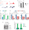
GATA6 directly activates the proepithelial transcription factors FOXA1 and FOXA2. (A) Representation of ChIP-Seq peaks on FOXA1 and FOXA2 promoters. (B) GATA6 binding to the promoters of FOXA1 and FOXA2 detected by ChIP-qPCR in PaTu8988S cells. (C–D) Expression of FOXA1 and FOXA2 in L3.6 (C), PaTu8988S and SK-PC-1 (D) cells infected with the indicated constructs, measured by RT-qPCR. (E) Expression of FOXA1 and FOXA2 proteins in GATA6-silenced PaTu8988S cells. Vinculin was used as loading control. (F) Luciferase-based reporter assay showing the activity of FOXA1 and FOXA2 promoter reporters in HEK293 cells transfected with empty vector (blue) or with vectors expressing either wild-type (light green) or mutated (dark green) GATA6. In all the panels, data are presented as mean±SEM of at least three independent experiments; *p<0.05, **p<0.01. ChIP-qPCR data are represented as % of input normalised against a negative control sequence, compared with binding of non-specific IgG; statistical significance is calculated for the enrichment of GATA6 binding to the region of interest, compared with the negative sequence.
Figure 5
Figure 5
GATA6 loss in human pancreatic ductal adenocarcinoma (PDAC) is associated with altered differentiation. (A) Expression of GATA6, FOXA2, E-cadherin and KRT14 in two PDAC samples, detected by immunohistochemistry. Top: cells retaining GATA6 expression are FOXA2high, E-cadherinhigh and KRT14neg; bottom: GATA6neg cells are FOXA2low, E-cadherinlow and KRT14pos. Scale bar: 50 μm. (B) Scatter plots showing correlated expression of GATA6, FOXA2 and E-cadherin mRNA in the PDAC meta-dataset. (C) Proportion of tumours showing GATA6 amplification (blue) or genomic loss (red) in the combined analysis of three PDAC gene copy number variation datasets. The percentage of GATA6 losses that were independent from loss of SMAD4 is represented in dark red.
Figure 6
Figure 6
GATA6 expression is associated with outcome and with response to adjuvant therapy. (A) Kaplan–Meier plot of the overall survival for patients included in the French series. (B) Median survival of patients included in the French series, classified according to GATA6 level. The value of p=0.003 calculated with Mann–Whitney U test. (C) Kaplan–Meier plot of the overall survival for patients included in the 5-fluorouracil (5-FU)/leucovorin arm of the ESPAC-3 trial. (D) Kaplan–Meier plot of the overall survival for patients included in the gemcitabine arm of the ESPAC-3 trial.
Figure 7
Figure 7
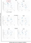
GATA6 expression negatively correlates with sensitivity to 5-fluorouracil (5-FU) in pancreatic ductal adenocarcinoma (PDAC) cells. Scatter plots showing cell survival upon treatment with the indicated doses of 5-FU (top), gemcitabine (middle) and paclitaxel (bottom), plotted against GATA6 protein level. Red square indicates significant correlation. Survival was normalised against DMSO-treated cells. Data are presented as the average value of at least three independent experiments.

References

    1. Siegel RL, Miller KD, Jemal A. Cancer statistics, 2015. CA Cancer J Clin 2015;65:5–29. 10.3322/caac.21254 - DOI - PubMed
    1. Ferlay J, Steliarova-Foucher E, Lortet-Tieulent J, et al. . Cancer incidence and mortality patterns in Europe: estimates for 40 countries in 2012. Eur J Cancer 2013;49:1374–403. 10.1016/j.ejca.2012.12.027 - DOI - PubMed
    1. Oettle H, Post S, Neuhaus P, et al. . Adjuvant chemotherapy with gemcitabine vs observation in patients undergoing curative-intent resection of pancreatic cancer: a randomized controlled trial. JAMA 2007;297:267–77. 10.1001/jama.297.3.267 - DOI - PubMed
    1. Neoptolemos JP, Dunn JA, Stocken DD, et al. . Adjuvant chemoradiotherapy and chemotherapy in resectable pancreatic cancer: a randomised controlled trial. Lancet 2001;358:1576–85. 10.1016/S0140-6736(01)06651-X - DOI - PubMed
    1. Neoptolemos JP, Stocken DD, Friess H, et al. . A randomized trial of chemoradiotherapy and chemotherapy after resection of pancreatic cancer. N Engl J Med 2004;350:1200–10. 10.1056/NEJMoa032295 - DOI - PubMed

Publication types

MeSH terms