. 2016 Jun 20:12:1744806916654144.
doi: 10.1177/1744806916654144. Print 2016.

Mycolactone-mediated neurite degeneration and functional effects in cultured human and rat DRG neurons: Mechanisms underlying hypoalgesia in Buruli ulcer

Affiliations

Mycolactone-mediated neurite degeneration and functional effects in cultured human and rat DRG neurons: Mechanisms underlying hypoalgesia in Buruli ulcer

U Anand et al. Mol Pain. .

Abstract

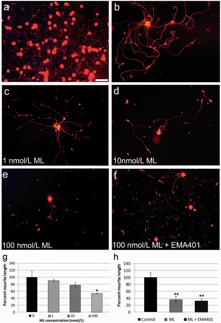
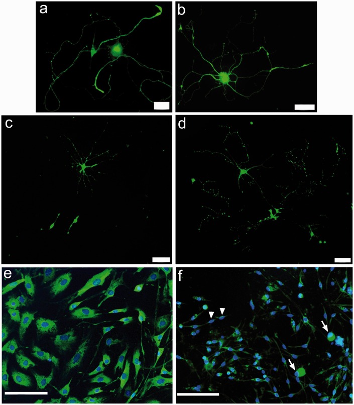
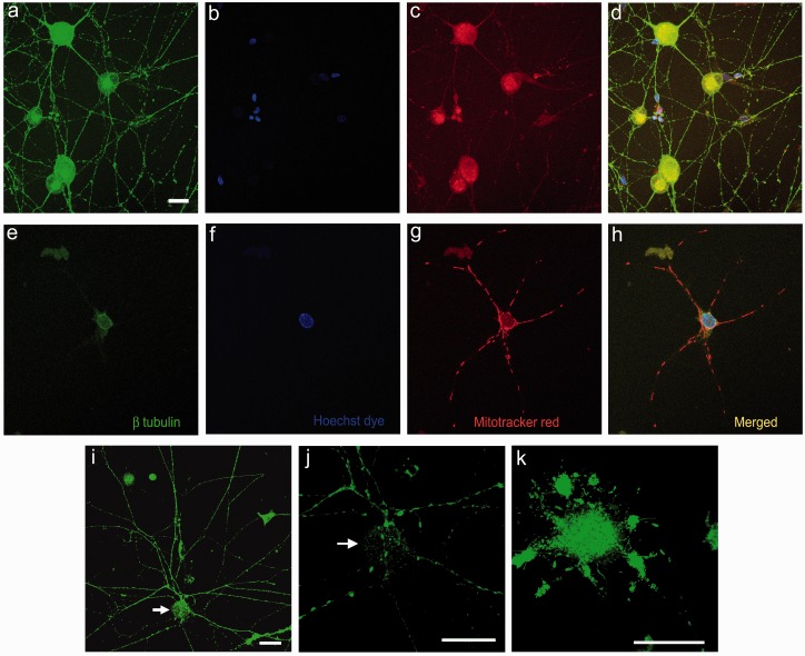
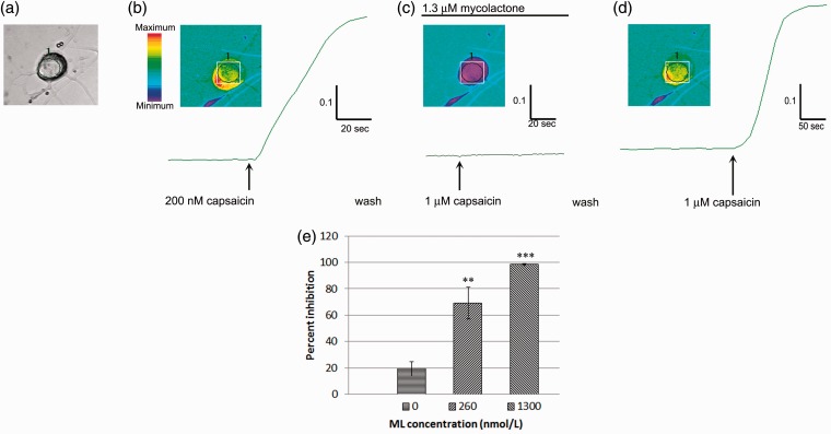
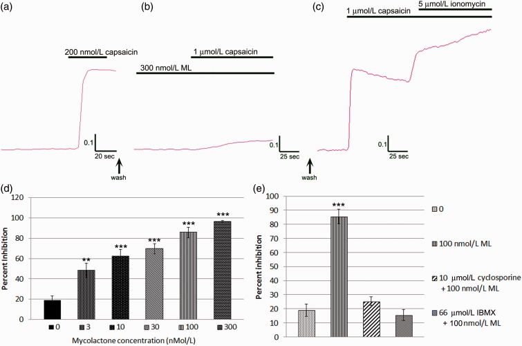
Background: Mycolactone is a polyketide toxin secreted by the mycobacterium Mycobacterium ulcerans, responsible for the extensive hypoalgesic skin lesions characteristic of patients with Buruli ulcer. A recent pre-clinical study proposed that mycolactone may produce analgesia via activation of the angiotensin II type 2 receptor (AT2R). In contrast, AT2R antagonist EMA401 has shown analgesic efficacy in animal models and clinical trials for neuropathic pain. We therefore investigated the morphological and functional effects of mycolactone in cultured human and rat dorsal root ganglia (DRG) neurons and the role of AT2R using EMA401. Primary sensory neurons were prepared from avulsed cervical human DRG and rat DRG; 24 h after plating, neurons were incubated for 24 to 96 h with synthetic mycolactone A/B, followed by immunostaining with antibodies to PGP9.5, Gap43, β tubulin, or Mitotracker dye staining. Acute functional effects were examined by measuring capsaicin responses with calcium imaging in DRG neuronal cultures treated with mycolactone.

Results: Morphological effects: Mycolactone-treated cultures showed dramatically reduced numbers of surviving neurons and non-neuronal cells, reduced Gap43 and β tubulin expression, degenerating neurites and reduced cell body diameter, compared with controls. Dose-related reduction of neurite length was observed in mycolactone-treated cultures. Mitochondria were distributed throughout the length of neurites and soma of control neurons, but clustered in the neurites and soma of mycolactone-treated neurons. Functional effects: Mycolactone-treated human and rat DRG neurons showed dose-related inhibition of capsaicin responses, which were reversed by calcineurin inhibitor cyclosporine and phosphodiesterase inhibitor 3-isobutyl-1-Methylxanthine, indicating involvement of cAMP/ATP reduction. The morphological and functional effects of mycolactone were not altered by Angiotensin II or AT2R antagonist EMA401.

Conclusion: Mycolactone induces toxic effects in DRG neurons, leading to impaired nociceptor function, neurite degeneration, and cell death, resembling the cutaneous hypoalgesia and nerve damage in individuals with M. Ulcerans infection.

Keywords: Buruli ulcer; TRPV1; apoptosis; calcium influx; hypoalgesia; mitochondria; mycolactone; neurite degeneration; neurons.


Figures

Figure 1.
Figure 1.
Loss of neurites in ML-treated rDRG neurons. PGP9.5 immunofluorescent neurons in control DRG cultures showing dense clusters (a) and a single neuron with profuse neurite outgrowth (b); Dose-related loss of neurites was observed after 24 h treatment with 1 nmol/L ML (c); 10 nmol/L ML (d); and 100 nmol/L ML (e); Co-incubation with the AT2R antagonist EMA401 did not affect ML-induced neurite degeneration (100 nmol/L each) (f); Graphs showing summary of neurite lengths after 24 h ML treatment (*p < 0.05, paired t-test) (g); Neurite lengths were significantly reduced after 48-h treatment with 100 nmol/L ML (**p = 0.010), and similar to neurons treated with combined EMA401 and ML (100 nmol/L each, **p = 0.007) (h). Bar in (a) = 50 m (same magnification in b–h).
Figure 2.
Figure 2.
Gap43 immunofluorescence was localized in the cell bodies and long profuse neurites in individual control rDRG neurons (a, b). Surviving neurons after 100 nmol/L ML treatment for 96 h showed shrunken cell bodies with disintegrated neurites (c, d). Non-neuronal cells comprising Schwann cells and fibroblasts in control cultures showed well-spread viable cells with distinct cytoplasm and nuclei (e). ML treatment resulted in shrunken neurons (arrows) and non-neuronal cells (arrowheads), with bright condensed nuclei typical of apoptosis (f). Bar in (a) and (c) =20 m, in (b) and (d) =50 m, and in (e) and (f) =100 m.
Figure 3.
Figure 3.
ML effect on rDRG neurons. Low power confocal images of control neurons (a–d) showing uniform distribution of β tubulin in the cell body and neurites (a), Hoechst dye stained nuclei (b), uniformly dispersed Mitotracker red labeled mitochondria (c) and the merged image with overlapping β tubulin and mitochondria (yellow, d). ML-treated neuron (100 nmol/L for 48 h) (e–h), showing loss of neurites and β tubulin (e), prominent nucleus (f), clumped Mitotracker Red positive mitochondria (g), and merged image (h). Live cell Mitotracker green labeling of mitochondria which are dispersed throughout the cell body (arrow) and neurites in a control neuron (i), and high power confocal images of the same neuron (j). In a ML-treated neuron, the Mitotracker label shows clumping of mitochondria restricted to the cell body, and loss of neurites (k). Bar in (a) =25 microns (a–h same magnification).
Figure 4.
Figure 4.
Acute effects of ML on capsaicin responses in hDRG neurons. Brightfield image of a human DRG neuron (a), showing increased fura 2 ratio in response to 200 nM capsaicin and the corresponding trace (b), followed by complete inhibition of 1 mol/L capsaicin response in the presence of 1.3 mol/L ML (c). The acute inhibitory effect of ML was reversible following washout, and the capsaicin response was restored (d). Graph showing percent inhibition of responses to 1 M capsaicin dose dependently inhibited in the presence of 260 nmol/L ML (**p = 0.003) and 1.3 mol/L ML**p = 0.004 (e).
Figure 5.
Figure 5.
Acute effects of ML on capsaicin responses in rDRG neurons. Trace showing increased fura2 ratio in response to 200 nM capsaicin (a). Near complete inhibition was observed in the presence of ML 300 nmol/L (b). The acute inhibitory effect of ML was reversed following a 30-min rest period after washout, and robust responses to capsaicin and ionomycin were observed (c). Graph showing ML dose-related inhibition of capsaicin responses following acute incubation with ML (d). Graph showing inhibition of capsaicin responses by 100 nmol/L ML, which were abolished in the presence of cyclosporine and IBMX (e). *p < 0.05, **p < 0.01, ***p < 0.001.

References

    1. WHO. Treatment of Mycobacterium Ulcerans Disease (Buruli Ulcer). Guidance for Health Workers, 2012; www.who.int/mediacentre/factsheets/fs199/en/ (accessed 1 June 2016).
    1. Einarsdottir T, Huygen K. Buruli ulcer. Hum Vaccines 2011; 7: 1198–1203. - PubMed
    1. Walsh DS, Prttaels F, Myers WM. Buruli ulcer: advances in understanding Mycobacterium Ulcerans infection. Dermatol Clin 2011; 29: 1–8. - PubMed
    1. Bozzo C, Tiberio R, Graziola F, et al. A Mycobacterium ulcerans toxin, mycolactone, induces apoptosis in primary human keratinocytes and in HaCaT cells. Microbes Infect 2010; 12: 1258–1263. - PubMed
    1. Gronberg A, Zettergren L, Bergh K, et al. Antioxidants protect keratinocytes against M. ulcerans mycolactone cytotoxicity. PloS One 2010; 5: e13839. - PMC - PubMed

Publication types

MeSH terms