. 2016 Nov 12;42(6):703-715.
doi: 10.1002/biof.1304. Epub 2016 Jun 21.

Pancreatic β-Cell production of CXCR3 ligands precedes diabetes onset

Affiliations

Pancreatic β-Cell production of CXCR3 ligands precedes diabetes onset

Susan J Burke et al. Biofactors. .

Abstract

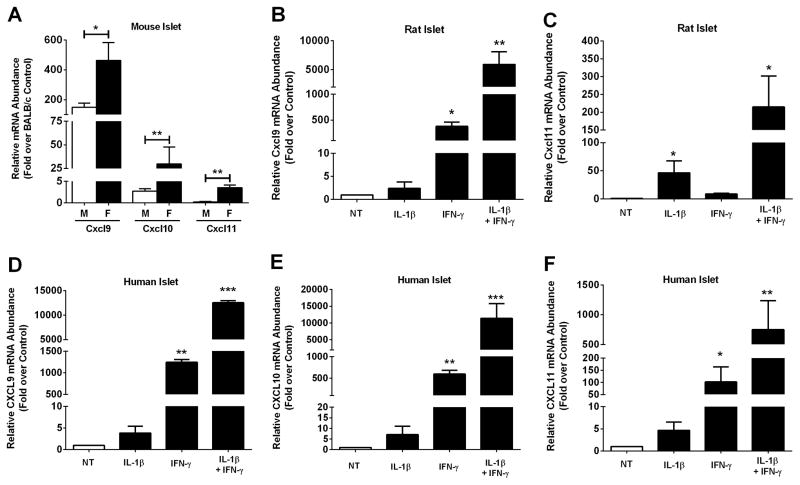
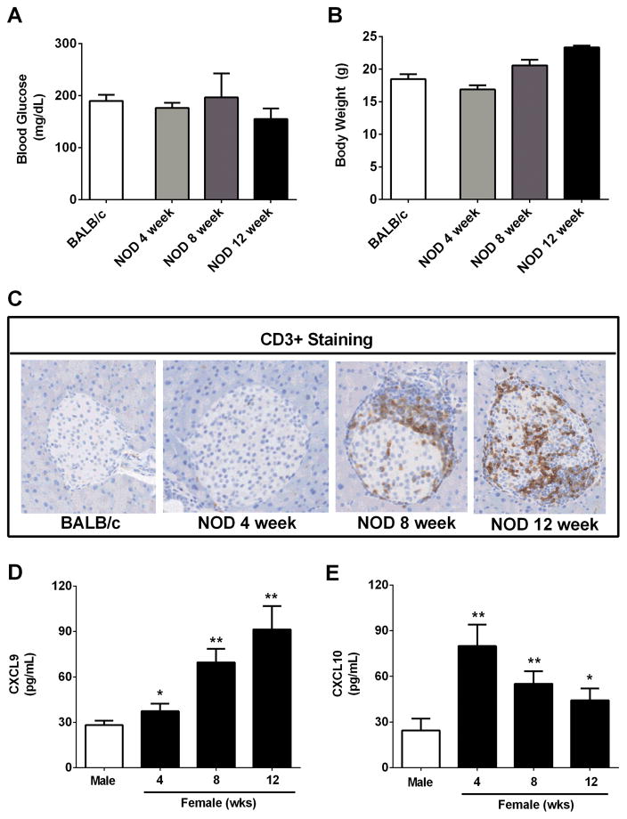
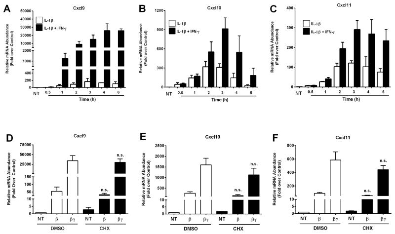
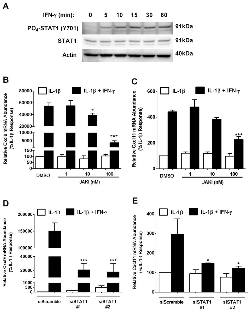
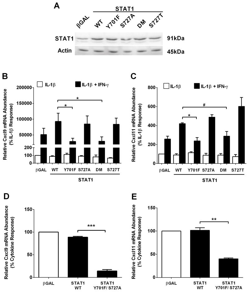
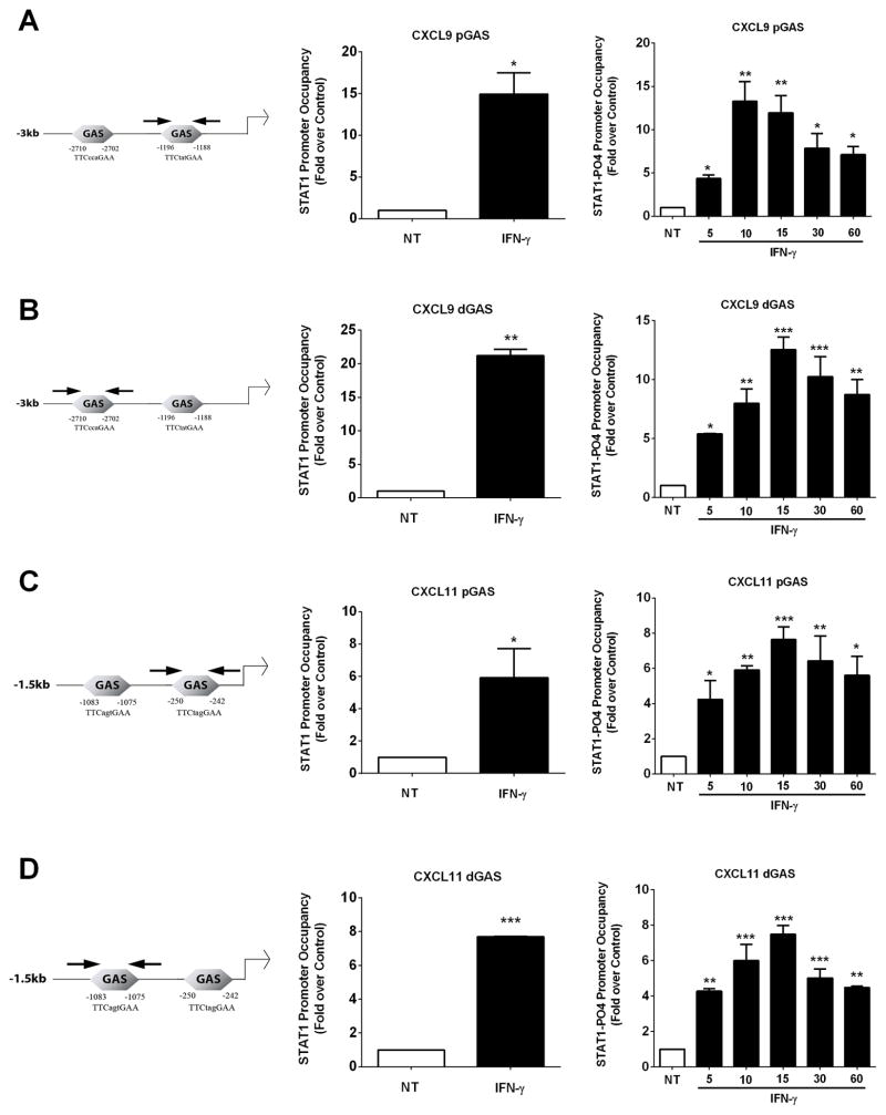
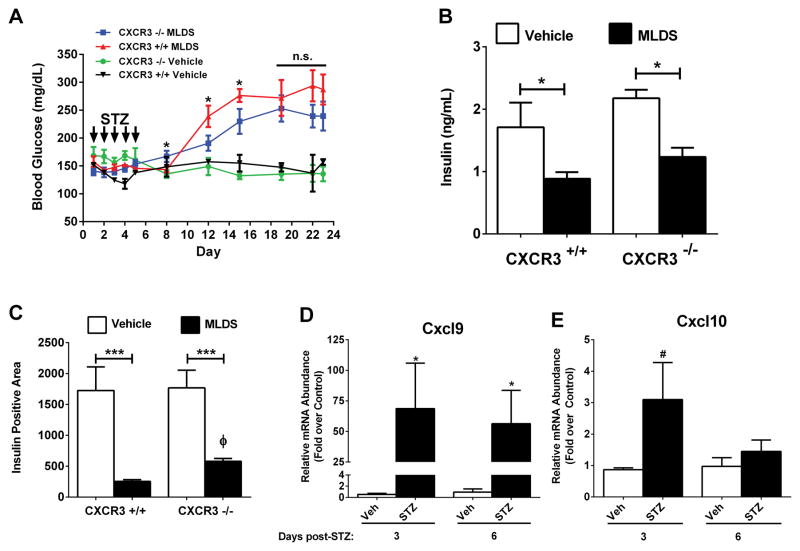
Type 1 diabetes mellitus (T1DM) results from immune cell-mediated reductions in function and mass of the insulin-producing β-cells within the pancreatic islets. While the initial trigger(s) that initiates the autoimmune process is unknown, there is a leukocytic infiltration that precedes islet β-cell death and dysfunction. Herein, we demonstrate that genes encoding the chemokines CXCL9, 10, and 11 are primary response genes in pancreatic β-cells and are also elevated as part of the inflammatory response in mouse, rat, and human islets. We further established that STAT1 participates in the transcriptional control of these genes in response to the pro-inflammatory cytokines IL-1β and IFN-γ. STAT1 is phosphorylated within five minutes after β-cell exposure to IFN-γ, with subsequent occupancy at proximal and distal response elements within the Cxcl9 and Cxcl11 gene promoters. This increase in STAT1 binding is coupled to the rapid appearance of chemokine transcript. Moreover, circulating levels of chemokines that activate CXCR3 are elevated in non-obese diabetic (NOD) mice, consistent with clinical findings in human diabetes. We also report herein that mice with genetic deletion of CXCR3 (receptor for ligands CXCL9, 10, and 11) exhibit a delay in diabetes development after being injected with multiple low doses of streptozotocin. Therefore, we conclude that production of CXCL9, 10, and 11 from islet β-cells controls leukocyte migration and activity into pancreatic tissue, which ultimately influences islet β-cell mass and function. © 2016 BioFactors, 42(6):703-715, 2016.

Keywords: autoimmunity; chemokine; diabetes; inflammation; islets; transcription.


Conflict of interest statement

The authors have no conflicts of interest to disclose.

Figures

Figure 1
Figure 1. The CXCR3 activating chemokines CXCL9, CXCL10, and CXCL11 are elevated in mouse, rat, and human islets during inflammation
A. Cxcl9, Cxcl10 and Cxcl11 transcript levels were measured in islets isolated from 10 week old male (n = 4) and female NOD mice (n = 5). Data are expressed relative to age-matched BALB/c control mice (n = 7). **p<0.01, *p<0.05. B–F. Islets from Wistar rats (B, C; n = 4 per group) or human islets (D–F; n =3 per group) were untreated (NT) or stimulated with 10 ng/mL IL-1β, 100 U/mL IFN-γ or both cytokines for 3 h. B–F. Relative mRNA abundance of CXCL9 (B, D), CXCL10 (E) and CXCL11 (C, F) was determined by RT-PCR. ***p<0.001 vs. NT, **p<0.01 vs. NT, *p<0.05 vs. NT.
Figure 2
Figure 2. Infiltration of T-lymphocytes into the pancreatic islets of NOD mice occurs prior to onset of hyperglycemia but is consistent with enhanced levels of CXCL9 and CXCL10 in serum
A–C. Blood glucose (A), body weight (B) and CD3+ staining (C) were assessed in 8 week old female BALB/c and 4, 8, and 12 week old female NOD mice. D,E. Serum CXCL9 (D) and CXCL10 (E) levels were determined in both male and female NOD mice (n = 4 mice per group). **p<0.01 vs. male, *p<0.05 vs. male.
Figure 3
Figure 3. Expression of the Cxcl9, Cxcl10 and Cxcl11 genes is increased in a cytokine-dependent manner in rat β-cell lines
A–C. 832/13 rat insulinoma cells were stimulated with 1 ng/mL IL-1β or IL-1β plus 100 U/mL IFN-γ for the indicated times (NT; no treatment). D–F. 832/13 cells were pretreated for 1 h with either DMSO or 0.5 μg/mL Cycloheximide (CHX). Cells were subsequently exposed to IL-1β (1 ng/mL) or the combination of IL-1β and IFN-γ (100 U/mL) for 2 h. Cellular mRNA levels of Cxcl9 (A, D), Cxcl10 (B, E) and Cxcl11 (C, F) were detected by RT-PCR. n.s. = not significant vs respective treatment in DMSO control group. Data are shown as means ± SEM from three independent experiments.
Figure 4
Figure 4. The JAK- STAT1 signaling pathway is required for cytokine-mediated activation of the Cxcl9 and Cxcl11 genes
A. 832/13 cells were exposed to 100 U/mL IFN-γ for the indicated times. PO4-STAT1Y701 and total STAT1 protein abundance were determined by immunoblotting. B, C. 832/13 cells were pre-treated for 1 h with increasing concentrations of JAKi (1 nM, 10 nM, 100 nM), followed by a 3 h stimulation with IL-1β alone (1 ng/mL) or IL-1β plus 100 U/mL IFN-γ. ***p<0.001 vs. DMSO (black bar), *p<0.05 vs. DMSO (black bar). D, E. 832/13 cells were transfected with two siRNA duplexes targeting STAT1 using a scrambled siRNA sequence duplex as a control. 48 h post- transfection cells were cultured for 3 h with 1 ng/ml IL-1β or IL-1β plus 100 U/ml IFN-γ. ***p<0.001 vs. siScramble (black bar), *p<0.05 vs. siScramble (black bar). Cxcl9 (B, D) and Cxcl11 (C, E) mRNA levels were quantified. Data are represented as means ± SEM from three independent experiments. The immunoblot in A was repeated on two separate occasions.
Figure 5
Figure 5. STAT1 phosphorylation at Y701, but not S727, is required for maximal cytokine-dependent activation of the Cxcl9 and Cxcl11 genes
A. 832/13 cells were transduced with adenoviruses encoding either βGAL, wild-type STAT1 (WT), STAT1Y701F, STAT1S727A, STAT1Y701F/S727A (DM; double mutant) or STAT1S727T. STAT1 abundance was determined by immunoblotting. B, C. 832/13 cells were transduced with the adenoviruses indicated in (A); 24 h post-transduction cells were stimulated for 3 h with either IL-1β (1 ng/mL) alone or IL-1β plus IFN-γ (100 U/mL). *p<0.05, #p<0.1. D, E. Rat islets were transduced with the indicated adenoviruses. 24 h post-transduction cells were stimulated with both IL-1β (10 ng/mL) and IFN-γ (100 U/mL) for 3 h. ***p<0.001, **p<0.01. Relative mRNA abundance of Cxcl9 (B, D) and Cxcl11 (C, E) was determined by RT-PCR. Date are expressed as means ± SEM from 3 (B, C) or 2 (D, E) individual experiments. The immunoblot in A was repeated on two individual occasions.
Figure 6
Figure 6. Phosphorylated STAT1 is recruited to the Cxcl9 and Cxcl11 promoters in response to IFN-γ
Schematic representation of distal and proximal GAS sites in the Cxcl9 and Cxcl11 promoters are shown (left panels). Arrows are the diagrammatic depiction of PCR amplicons. A–D. 832/13 cells were stimulated with 100 U/mL IFN-γ for either 20 mins (middle panels) or a time course (right panels). ChIP assays were performed to determine relative occupancy of total STAT1 (middle panels) and PO4-STAT1Y701 (right panels) on the Cxcl9 proximal (A) and distal promoter (B), and on the Cxcl11 proximal (C) and distal (D) promoter. ***p<0.001 vs. NT, **p<0.01 vs. NT, *p<0.05 vs. NT. Data are expressed as means ± SEM from 3–4 individual experiments.
Figure 7
Figure 7. Genetic Deletion of CXCR3 Delays Onset of Hyperglycemia Induced by Multiple Low Doses of Streptozotocin
A. Blood glucose levels in control (CXCR3+/+) and CXCR3 knockout (CXCR3−/−) mice were monitored for 23 days after starting the MLDS protocol (n = 9 per group). *p<0.05 vs. CXCR3−/− on respective days. B. Serum insulin levels were measured 23 days after initiation of MLDS protocol. C. Insulin positive area from formalin-fixed, paraffin-embedded pancreatic tissue was quantified (n = 9 per group). D, E. C57BL/6J mice were injected with STZ for 5 consecutive days. Islets were isolated at 3 and 6 days following the last injection, and transcript levels of Cxcl9 (D) and Cxcl10 (E) were determined by qPCR. ***p<0.001 (C), *p<0.05 (D), #p<0.1 (E), Φ, p<0.05 vs. CXCR3+/+ (C).

References

    1. Meier JJ. Beta cell mass in diabetes: a realistic therapeutic target? Diabetologia. 2008;51:703–713. - PubMed
    1. Tuomi T, Santoro N, Caprio S, Cai M, Weng J, Groop L. The many faces of diabetes: a disease with increasing heterogeneity. Lancet. 2014;383:1084–1094. - PubMed
    1. von Herrath MG, Oldstone MB. Interferon-gamma is essential for destruction of beta cells and development of insulin-dependent diabetes mellitus. J Exp Med. 1997;185:531–539. - PMC - PubMed
    1. Arnush M, Heitmeier MR, Scarim AL, Marino MH, Manning PT, Corbett JA. IL-1 produced and released endogenously within human islets inhibits beta cell function. J Clin Invest. 1998;102:516–526. - PMC - PubMed
    1. Sarkar SA, Lee CE, Victorino F, Nguyen TT, Walters JA, Burrack A, Eberlein J, Hildemann SK, Homann D. Expression and regulation of chemokines in murine and human type 1 diabetes. Diabetes. 2012;61:436–446. - PMC - PubMed

MeSH terms