. 2016 Jul 5;113(27):7632-7.
doi: 10.1073/pnas.1524907113. Epub 2016 Jun 20.

Pubertal activation of estrogen receptor α in the medial amygdala is essential for the full expression of male social behavior in mice

Affiliations

Pubertal activation of estrogen receptor α in the medial amygdala is essential for the full expression of male social behavior in mice

Kazuhiro Sano et al. Proc Natl Acad Sci U S A. .

Abstract

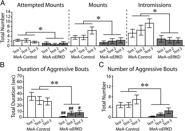
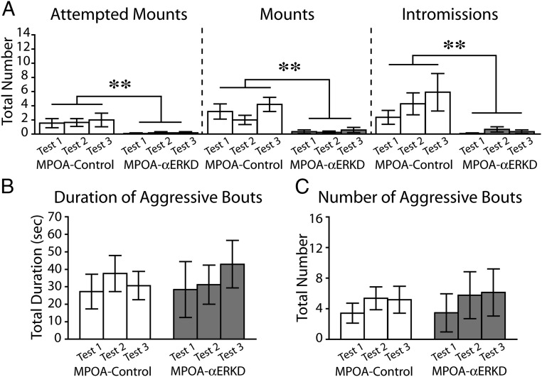
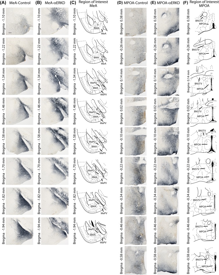
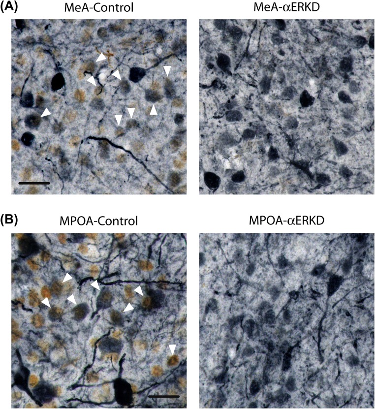
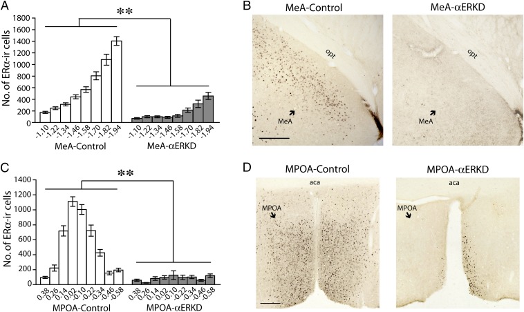
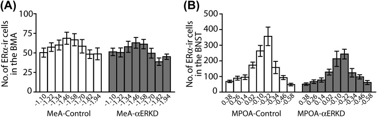
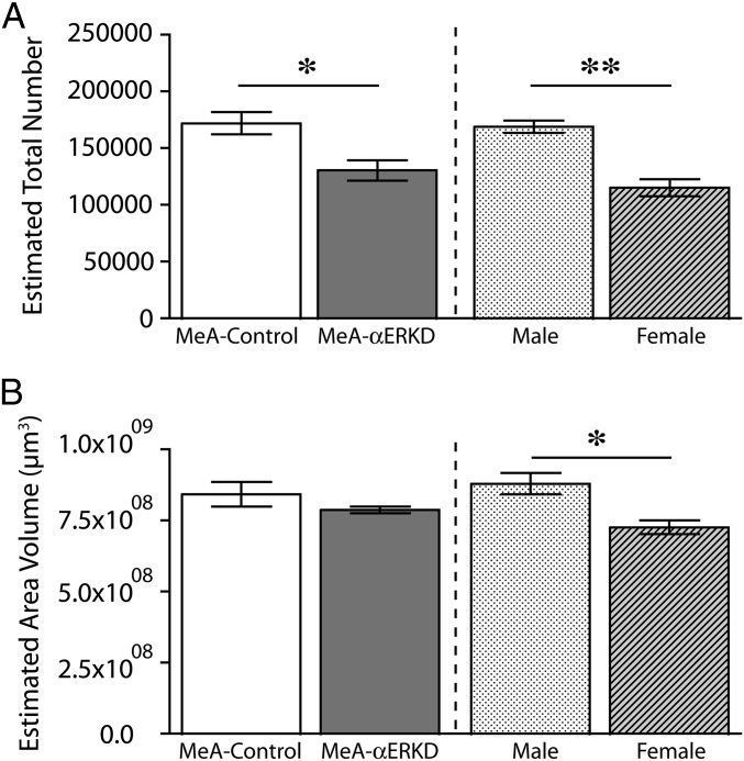
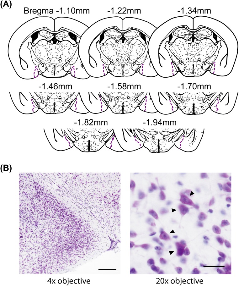
Testosterone plays a central role in the facilitation of male-type social behaviors, such as sexual and aggressive behaviors, and the development of their neural bases in male mice. The action of testosterone via estrogen receptor (ER) α, after being aromatized to estradiol, has been suggested to be crucial for the full expression of these behaviors. We previously reported that silencing of ERα in adult male mice with the use of a virally mediated RNAi method in the medial preoptic area (MPOA) greatly reduced sexual behaviors without affecting aggressive behaviors whereas that in the medial amygdala (MeA) had no effect on either behavior. It is well accepted that testosterone stimulation during the pubertal period is necessary for the full expression of male-type social behaviors. However, it is still not known whether, and in which brain region, ERα is involved in this developmental effect of testosterone. In this study, we knocked down ERα in the MeA or MPOA in gonadally intact male mice at the age of 21 d and examined its effects on the sexual and aggressive behaviors later in adulthood. We found that the prepubertal knockdown of ERα in the MeA reduced both sexual and aggressive behaviors whereas that in the MPOA reduced only sexual, but not aggressive, behavior. Furthermore, the number of MeA neurons was reduced by prepubertal knockdown of ERα. These results indicate that ERα activation in the MeA during the pubertal period is crucial for male mice to fully express their male-type social behaviors in adulthood.

Keywords: estrogen receptor α; medial amygdala; pubertal period; social behavioral network; testosterone.


Conflict of interest statement

The authors declare no conflict of interest.

Figures

Fig. 1.
Fig. 1.
Effects of prepubertal ERα knockdown in the MeA on male sexual and aggressive behaviors. (A) Number of attempted mounts (Left), mounts (Middle), and intromissions (Right) were significantly reduced in the MeA-αERKD compared with the MeA-control group. (B and C) Both the duration and number of aggressive bouts were significantly reduced in the MeA-αERKD compared with the MeA-control group. **P < 0.01, *P < 0.05, as indicated in the figures. ##P < 0.01, #P < 0.05 vs. MeA-control group in the respective test. Data are presented as mean ± SEM.
Fig. 2.
Fig. 2.
Effects of prepubertal ERα knockdown in the MPOA on male sexual and aggressive behaviors. (A) Number of attempted mounts (Left), mounts (Middle), and intromissions (Right) were significantly reduced in the MPOA-αERKD compared with the MPOA-control group. (B and C) There was no difference between the MPOA-control and MPOA-αERKD groups in either (B) duration or (C) number of aggressive bouts. **P < 0.01. Data are presented as mean ± SEM.
Fig. S1.
Fig. S1.
GFP distribution patterns throughout the rostral-caudal extent of the target regions. (A and B) Representative photomicrographs of ERα-GFP double-labeled sections of MeA-control and MeA-αERKD mice, and (C) histological diagrams of the MeA (defined as the area combined MePD and MePV) throughout the rostral-caudal extent (Bregma −1.10 to −1.94 with 120-µm intervals). (D and E) Representative photomicrographs of ERα-GFP double-labeled sections of MPOA-control and MPOA-αERKD mice, and (F) histological diagrams of the MPOA throughout the rostral-caudal extent (Bregma 0.38 to −0.58 with 120-µm intervals). (Scale bars: 200 µm.) BMA, basomedial amygdala; BNSTV, bed nucleus of the stria terminalis, ventral; MePD, medial amygdala, posterodorsal; MePV, medial amygdala, posteroventral; MPOA, medial preoptic area.
Fig. S2.
Fig. S2.
Double-label immunohistochemistry for ERα (brown) and GFP (blue-gray). (A) Representative photomicrographs of double-labeled MeA sections (Bregma −1.67) of MeA-control and MeA-αERKD. (B) Representative photomicrographs of double-labeled MPOA sections (Bregma −0.07) of MPOA-control and MPOA-αERKD. The absence of ERα-GFP double-labeled cells in the MeA-αERKD or MPOA-αERKD sections indicates that AAV-shERα used in the present study successfully knocked down ERα expression in the transfected cells. (Scale bars: 20 µm.) The white arrowheads indicate ERα-GFP double-labeled cells.
Fig. 3.
Fig. 3.
Immunohistochemical evaluations of ERα knockdown in the MeA and MPOA. (A and C) The number of ERα-immunoreactive cells in the target regions was significantly reduced in the αERKD groups compared with their respective control groups throughout the rostrocaudal axis (A) in the MeA (Bregma −1.10 to −1.94; defined as the area combined MePD and MePV) (Fig. S1C) and (C) in the MPOA (Bregma 0.38 to −0.58) (Fig. S1F). **P < 0.01. Data are presented as mean ± SEM. (B and D) Representative photomicrographs of ERα-immunoreactive cells in brain sections from (B) the MeA-control and MeA-αERKD groups and (D) the MPOA-control and MPOA-αERKD groups. (Scale bars: 200 µm.)
Fig. S3.
Fig. S3.
Confirmation of site specificity of ERα knockdown. (A) Number of ERα-immunoreactive cells in the BMA, the lateral adjacent area to the MeA, was not different between the MeA-αERKD and MeA-control groups throughout the rostrocaudal axis (Bregma −1.10 to −1.94) (see Fig. S1C for the definition of the BMA). (B) Number of ERα-immunoreactive cells in the ventral portion of the BNST, the dorsal adjacent area to the MPOA, was not different between the MPOA-αERKD and MPOA-control groups throughout the rostrocaudal axis (Bregma 0.38 to −0.58) (see Fig. S1F for the definition of the ventral portion of the BNST). All data are presented as mean ± SEM.
Fig. 4.
Fig. 4.
Effects of prepubertal ERα knockdown on the morphology of the MeA examined in adults. (A) The number of neuronal cells in the MeA was significantly reduced in the MeA-αERKD compared with the MeA-control (Left). In uninjected reference groups, gonadally intact males had a significantly greater number of neuronal cells in the MeA compared with gonadally intact females (Right). (B) There was no difference between the MeA-control and MeA-αERKD groups in the regional volume of the MeA (Left) whereas gonadally intact males had greater regional volume compared with gonadally intact females (Right). **P < 0.01, *P < 0.05, as indicated. Data are presented as mean ± SEM.
Fig. S4.
Fig. S4.
(A) Histological diagrams of the MeA included in the morphological evaluation. (B) Representative photomicrographs of a Nissl-stained MeA section viewed under 4× objective (Left) and the same section viewed under 20× objective (Right). (Scale bars: Left, 200 µm; Right, 20 µm.) The black arrowheads indicate neuronal cells.
Fig. S5.
Fig. S5.
Results of a supplemental experiment performed to confirm successful vector uptake and ERα knockdown before the onset of puberty in mice unilaterally injected with AAV-shERα vectors on PND 21. (A) Percentage of NeuN and GFP double-labeled (NeuN+ and GFP+) cells in the total number of NeuN+ (Left) or GFP+ (Right) cells. About 90% of NeuN+ cells in the MPOA also expressed GFP at the time point of 5, 10, or 15 d after AAV vector injection. All data are presented as mean of two mice. N/A, not applicable due to absence of GFP expression. (B) Representative photomicrographs of a NeuN-GFP double-labeled section in the injected side of the MPOA at the time point of 15 d after vector injection. (Scale bar: 200 µm.) (C) Representative photomicrographs demonstrating immunohistochemical labeling for GFP in the MPOA sections (Top) and that for ERα in the adjacent sections (Bottom) at four different time points (4 h and on 5, 10, or 15 d) after unilateral injection of AAV-shERα. At 4 h, GFP expression was not detected in the injected side (left hemisphere), and the expression of ERα was not affected. On 5, 10, and 15 d after injection, GFP-immunopositive cells were detected, and expression of ERα were greatly reduced in the injected side. These results suggest that AAV-shERα used in the present study successfully knocked down ERα expression in the targeted regions by the fifth day after injection: i.e., PND 26, before the onset of puberty. (Scale bar: 200 µm.)

References

    1. Ogawa S, Lubahn DB, Korach KS, Pfaff DW. Behavioral effects of estrogen receptor gene disruption in male mice. Proc Natl Acad Sci USA. 1997;94(4):1476–1481. - PMC - PubMed
    1. Ogawa S, et al. Modifications of testosterone-dependent behaviors by estrogen receptor-α gene disruption in male mice. Endocrinology. 1998;139(12):5058–5069. - PubMed
    1. Ogawa S, et al. Abolition of male sexual behaviors in mice lacking estrogen receptors α and β (α β ERKO) Proc Natl Acad Sci USA. 2000;97(26):14737–14741. - PMC - PubMed
    1. Toda K, Saibara T, Okada T, Onishi S, Shizuta Y. A loss of aggressive behaviour and its reinstatement by oestrogen in mice lacking the aromatase gene (Cyp19) J Endocrinol. 2001;168(2):217–220. - PubMed
    1. Toda K, et al. Oestrogen at the neonatal stage is critical for the reproductive ability of male mice as revealed by supplementation with 17β-oestradiol to aromatase gene (Cyp19) knockout mice. J Endocrinol. 2001;168(3):455–463. - PubMed

Publication types

Substances