. 2016 Jun 28;113(26):7154-9.
doi: 10.1073/pnas.1603827113. Epub 2016 Jun 20.

Structure of a phleboviral envelope glycoprotein reveals a consolidated model of membrane fusion

Affiliations

Structure of a phleboviral envelope glycoprotein reveals a consolidated model of membrane fusion

Steinar Halldorsson et al. Proc Natl Acad Sci U S A. .

Abstract

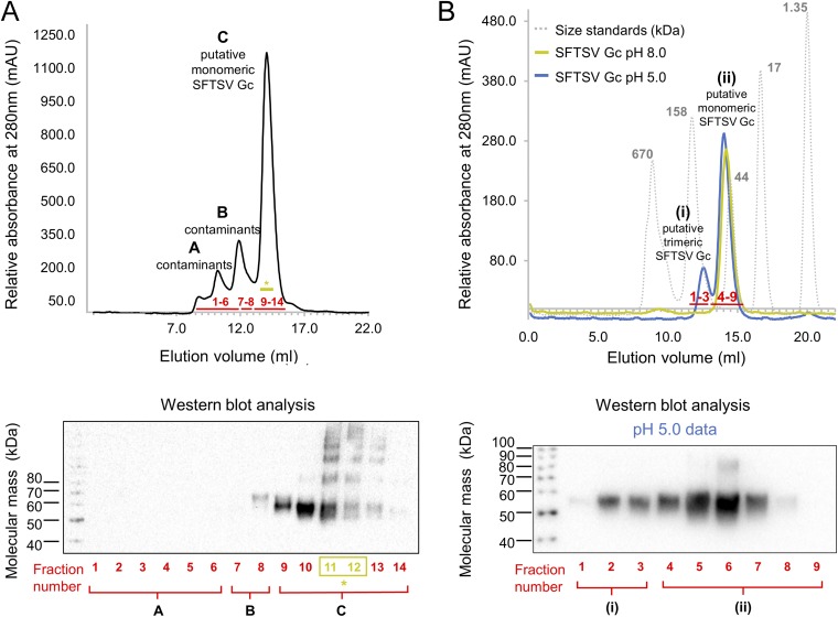
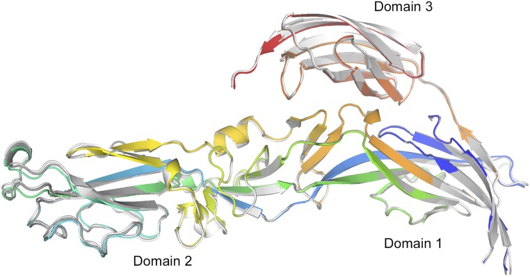
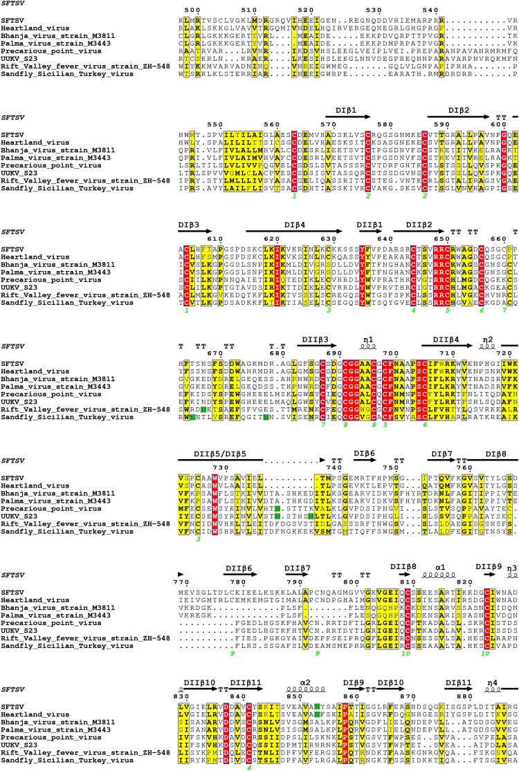
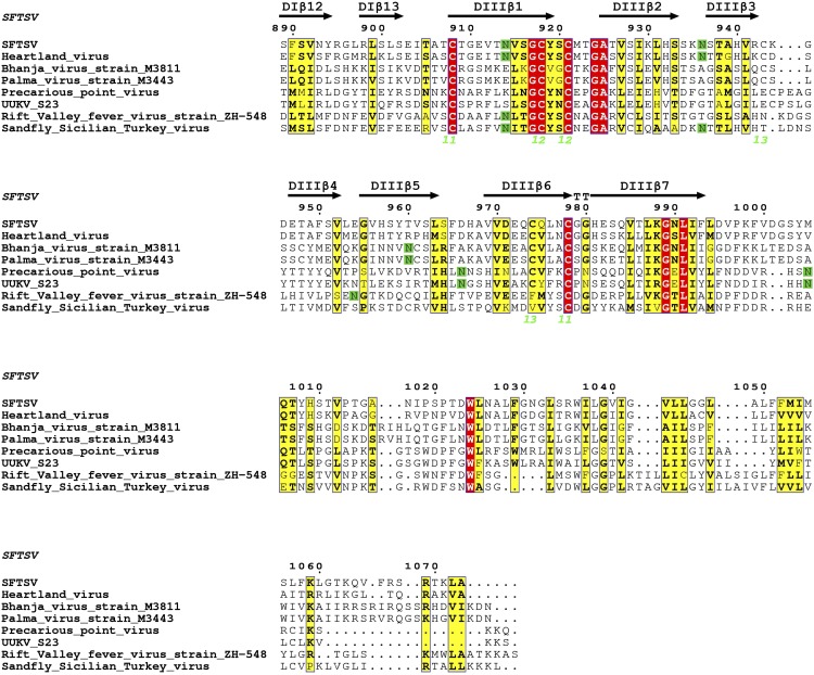
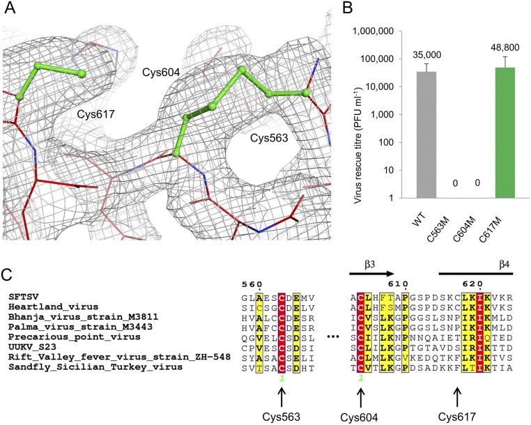
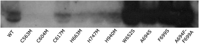
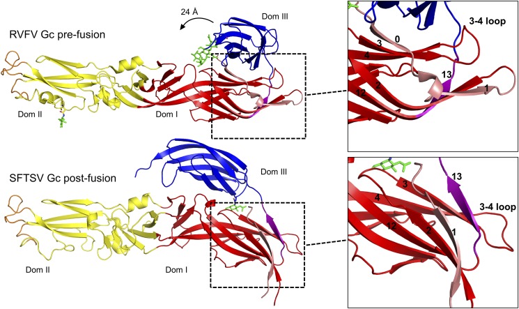
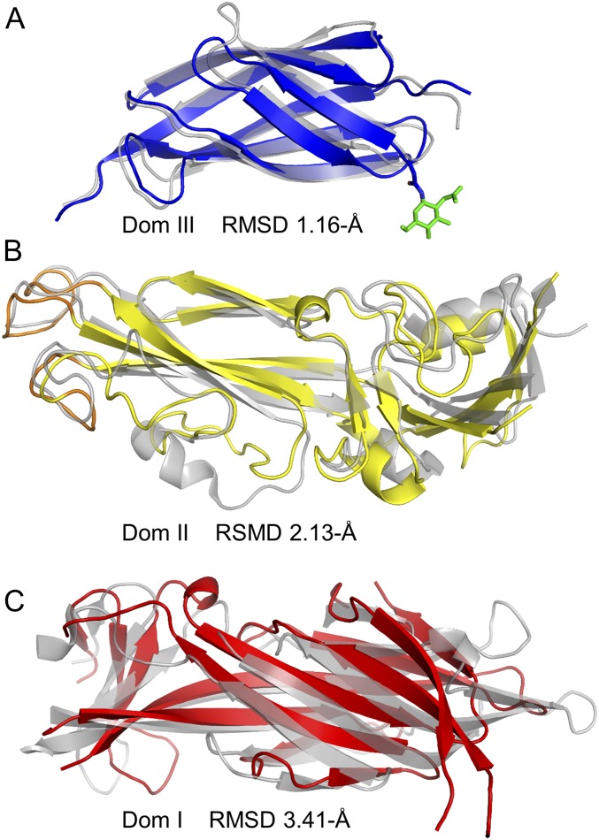
An emergent viral pathogen termed severe fever with thrombocytopenia syndrome virus (SFTSV) is responsible for thousands of clinical cases and associated fatalities in China, Japan, and South Korea. Akin to other phleboviruses, SFTSV relies on a viral glycoprotein, Gc, to catalyze the merger of endosomal host and viral membranes during cell entry. Here, we describe the postfusion structure of SFTSV Gc, revealing that the molecular transformations the phleboviral Gc undergoes upon host cell entry are conserved with otherwise unrelated alpha- and flaviviruses. By comparison of SFTSV Gc with that of the prefusion structure of the related Rift Valley fever virus, we show that these changes involve refolding of the protein into a trimeric state. Reverse genetics and rescue of site-directed histidine mutants enabled localization of histidines likely to be important for triggering this pH-dependent process. These data provide structural and functional evidence that the mechanism of phlebovirus-host cell fusion is conserved among genetically and patho-physiologically distinct viral pathogens.

Keywords: bunyavirus; emerging virus; phlebovirus; structure; viral membrane fusion.


Conflict of interest statement

The authors declare no conflict of interest.

Figures

Fig. 1.
Fig. 1.
Crystal structure of SFTSV Gc in the postfusion conformation. (A) Domain diagram of the full M segment of SFTSV containing Gn and Gc with the crystallized ectodomain colored by domain: domain I in red, domain II in yellow, and domain III in blue. (B) SFTSV Gc in the postfusion trimeric conformation. The full trimer is shown on Left in cartoon representation and is colored according to domain as in A. Glycans observed in the crystal structure are shown as green sticks. On Right, a single protomer is shown in cartoon representation with the remainder of the trimer shown as a white van der Waals surface.
Fig. S1.
Fig. S1.
Size exclusion (SEC) and Western blot analysis of purified SFTSV Gc. SEC was performed on a Superdex Increase 200 10/30 column (Amersham) equilibrated in 150 mM NaCl and either 10 mM Tris·HCl, pH 8.0, or 20 mM citrate–NaOH, pH 5.0 (see SI Materials and Methods). (A) SEC of SFTSV Gc run at pH 8.0 following immobilized nickel affinity purification revealed one major peak (peak C) and two minor peaks (peaks A and B). Western blot analysis of fractions confirmed that peaks A and B contained contaminants from recombinant protein expression and peak C contained hexa-histidine–tagged SFTSV Gc. Fractions colored in olive green (*) were pooled and used for further SEC analysis in B. (B) Peak fractions 11 and 12 from SEC performed in A were split in two and run at pH 8.0 and pH 5.0. A gel filtration standard (Bio-Rad, gray) was run on the same column for comparison. At pH 8.0 (green), a single peak (ii) corresponding to putative monomeric SFTSV Gc was observed. At pH 5.0 (blue), a second small peak (i), corresponding to a putative trimer, was observed in addition to peak ii. Western blot analysis of pH 5.0 SEC fractions revealed that both the monomeric (ii) and putative trimeric (i) peaks were composed of SFTSV Gc, indicating that exposure to acidic environments may trigger SFTSV Gc trimerization in solution.
Fig. S2.
Fig. S2.
Structure and fold of SFTSV Gc protomer. An overlay of the three SFTSV Gc chains observed in the asymmetric unit is shown. One protomer of SFVTSV Gc is shown in cartoon representation and is colored as a rainbow ramped from blue (N terminus) to red (C terminus), and the other two overlaid protomers are shown in gray. The average rmsd is 0.64 Å over 428 Cα residues.
Fig. S3.
Fig. S3.
Sequence alignment of phleboviral Gc glycoproteins of selected phleboviruses, numbered according to SFTSV. Sequences of eight phleboviral M segments from four distinct clades were chosen and aligned (see Materials and Methods). The alignment numbering is based on SFTSV M segment. Fully conserved residues are shown in red and partially conserved residues in yellow. Residues highlighted green are predicted to be glycosylated; disulfide bonds observed in the SFTSV Gc crystal structure are numbered below the sequence in lime green. Secondary structure elements are labeled above the alignment, with β-strands shown as arrows and with helices (α-helix, α; 310 helix η) shown as spirals.
Fig. S3.
Fig. S3.
Sequence alignment of phleboviral Gc glycoproteins of selected phleboviruses, numbered according to SFTSV. Sequences of eight phleboviral M segments from four distinct clades were chosen and aligned (see Materials and Methods). The alignment numbering is based on SFTSV M segment. Fully conserved residues are shown in red and partially conserved residues in yellow. Residues highlighted green are predicted to be glycosylated; disulfide bonds observed in the SFTSV Gc crystal structure are numbered below the sequence in lime green. Secondary structure elements are labeled above the alignment, with β-strands shown as arrows and with helices (α-helix, α; 310 helix η) shown as spirals.
Fig. S4.
Fig. S4.
Aberrant Cys617 is found solely in SFTSV and Heartland virus. (A) Electron density (2Fo-Fc contoured at 1.0 σ, gray) around cysteines Cys563, Cys604, and Cys617 in SFTSV Gc. (B) SFTSV encoding single cysteine mutations (C563M, C604M, and C617M) were derived by reverse genetics, and the titers of these recombinant viruses, measured in PFUs, were compared with that of WT SFTSV. Only the C617M mutants were rescued and titrated to levels comparable with the WT virus, whereas C563M and C604M failed to rescue. (C) Sequence alignment of selected phleboviruses (colored and annotated as in Fig. S3) reveals that Cys563 and Cys604 are conserved among all selected phleboviruses, but Cys617 is only found in the closely related Heartland virus.
Fig. S5.
Fig. S5.
Western blot analysis confirms the secreted and soluble expression of SFTSV Gc ectodomain mutants in mammalian HEK 293T cells (see SI Materials and Methods). The WT SFTSV Gc ectodomain construct crystallized in this manuscript was used as a control. Two mutants, C563M and C604M, were not secreted and folded. All other mutants were expressed at levels equivalent to or greater than WT. Fusion loop mutants W652S, A694S, F966S, and A694F-F699A exhibited a greater level of overall expression with respect to WT, which may possibly be attributed to the increased protein stability achieved by the removal of solvent-exposed hydrophobic amino acids in the putative fusion loops.
Fig. 2.
Fig. 2.
Structural rearrangements of phleboviral Gc from prefusion to postfusion conformations. Single protomers of RVFV Gc (PDB ID code 4HJ1) and SFTSV Gc are shown in cartoon representation and colored as in Fig. 1. Glycans are shown as green sticks. Zoom-in panels of domain I are shown on the right side and highlight the strand swap occurring between pre- and postfusion states. In the postfusion conformation, strand 13 (purple) reorientates around the 3–4 loop, forming a β-sheet with strands 3 and 4, and strand 0 (pink) becomes continuous with strand 1 (pink).
Fig. S6.
Fig. S6.
Overlay analysis of individual SFTSV Gc and RVFV Gc domains. Structures are shown as cartoons, with RVFV Gc colored gray and SFTSV Gc colored according to domain boundaries (domain I, red; domain II, yellow; and domain III, blue). Calculated rmsd values are shown. Overlay of (A) domain III (1.16 Å rmsd over 81 Cα residues), (B) domain II (2.13 Å rmsd of over 117 Cα residues), and (C) domain I (3.41 Å rmsd over 109 Cα residues).
Fig. 3.
Fig. 3.
The putative fusion loops of SFTSV Gc are conformationally rigid and contain functionally essential residues. (A) An overlay of the fusion loops of SFTSV Gc (colored as in Fig. 1) and RVFV Gc (gray; PDB ID code 4HJ1) reveals highly similar conformations. Side chains from hydrophobic amino acids from SFTSV Gc are shown as orange sticks. Disulphide bonds are shown as green sticks. (B) Sequence alignment of fusion loops across selected phleboviruses. Residues highlighted red are fully conserved and yellow are partially conserved. Residues tested by site-directed mutagenesis are highlighted by arrows. Phe699 is fully conserved, whereas Trp652 is more varied among phleboviral sequences (SI Materials and Methods). (C) SFTSV encoding single and double site-directed mutations at the putative fusion loops were derived by reverse genetics, and the titers of these recombinant viruses, measured in plaque forming units (PFUs), were compared with wild-type (WT) SFTSV.
Fig. S7.
Fig. S7.
Electron density of SFTSV Gc glycans. Three N-linked glycosylation sites are predicted for SFTSV Gc: Asn853, Asn914, and Asn936. The 2Fo-Fc map is shown in gray at 1.0 σ, and only the glycan of Asn936 could be built with confidence.
Fig. S8.
Fig. S8.
Glycan analysis of SFTSV Gc. Enzymatically released glycans of the recombinantly expressed ectodomain of SFTSV Gc, produced in the absence of glycosylation inhibitors, were fluorescently labeled and probed by HILIC-UPLC analysis. (Upper) The overall glycan profile before (black) and after (magenta) endoglycosidase H digestion. Negligible levels of hybrid- and oligomannose-type glycans are revealed, with the spectrum being dominated by processed, complex-type glycans. (Lower) The glycan profile before (black) and after (blue) disialylation, highlighting a significant abundance of sialic acid-containing N-linked glycans.
Fig. S9.
Fig. S9.
Position of glycosylation sites from selected phleboviruses mapped onto the crystal structure of SFTSV Gc. Predicted glycosylation sites of the eight selected phleboviruses (Fig. S3) were analyzed and placed on equivalent sites on SFTSV Gc as spheres. The site numbering corresponds to the SFTSV Gc amino acid sequence. Glycosylation sites belonging to SFTSV Gc are shown in green, and sites of other viruses are shown in gray.
Fig. 4.
Fig. 4.
Surface-exposed His663, His747, and His940 on SFTSV Gc domains I and III play integral roles in the virus life cycle. (A) A single SFTSV Gc protomer is shown in cartoon representation (colored as in Fig. 1) with the remainder of the trimer shown as a white van der Waals surface. Selected surface-exposed histidines are shown as purple sticks. Zoom-in panels highlight the location of these residues within each of the three domains, and residues surrounding His747 and His940 from adjacent protomers are shown as white sticks. (B) SFTSV encoding single mutations of His663, His747, and His940 were derived by reverse genetics, and the titers of these recombinant viruses, measured in PFUs, were compared with that of WT SFTSV.

Similar articles

Cited by

References

    1. Yu X-J, et al. Fever with thrombocytopenia associated with a novel bunyavirus in China. N Engl J Med. 2011;364(16):1523–1532. - PMC - PubMed
    1. Cui N, et al. Severe fever with thrombocytopenia syndrome bunyavirus-related human encephalitis. J Infect. 2015;70(1):52–59. - PubMed
    1. Ding F, et al. Epidemiologic features of severe fever with thrombocytopenia syndrome in China, 2011-2012. Clin Infect Dis. 2013;56(11):1682–1683. - PubMed
    1. Lam TT-Y, et al. Evolutionary and molecular analysis of the emergent severe fever with thrombocytopenia syndrome virus. Epidemics. 2013;5(1):1–10. - PMC - PubMed
    1. Zhang L, et al. Antibodies against severe fever with thrombocytopenia syndrome virus in healthy persons, China, 2013. Emerg Infect Dis. 2014;20(8):1355–1357. - PMC - PubMed

Publication types

Substances

Associated data

LinkOut - more resources