

Case Reports
doi: 10.2484/rcr.v7i3.708.
eCollection 2012.
Plexiform neurofibroma in a 16-year-old girl
- PMID: 27326300
- PMCID: PMC4899673
- DOI: 10.2484/rcr.v7i3.708
Item in Clipboard
Case Reports
Plexiform neurofibroma in a 16-year-old girl
Radiol Case Rep.
.
Abstract
Plexiform neurofibroma is a rare, poorly defined benign tumor of the peripheral nerve sheath. It spreads out just under the skin, or deeper in the body, and occurs exclusively in patients with neurofibromatosis type I. Facial plexiform neurofibroma may produce various degrees of cosmetic and functional deformities in the head and neck region. It is a virtually pathognomonic and often disabling feature of neurofibromatosis type I. We present a case of plexiform neurofibroma in an 18-year-old female.
Keywords: CT, computed tomography; MRI, magnetic resonance imaging.
Figures
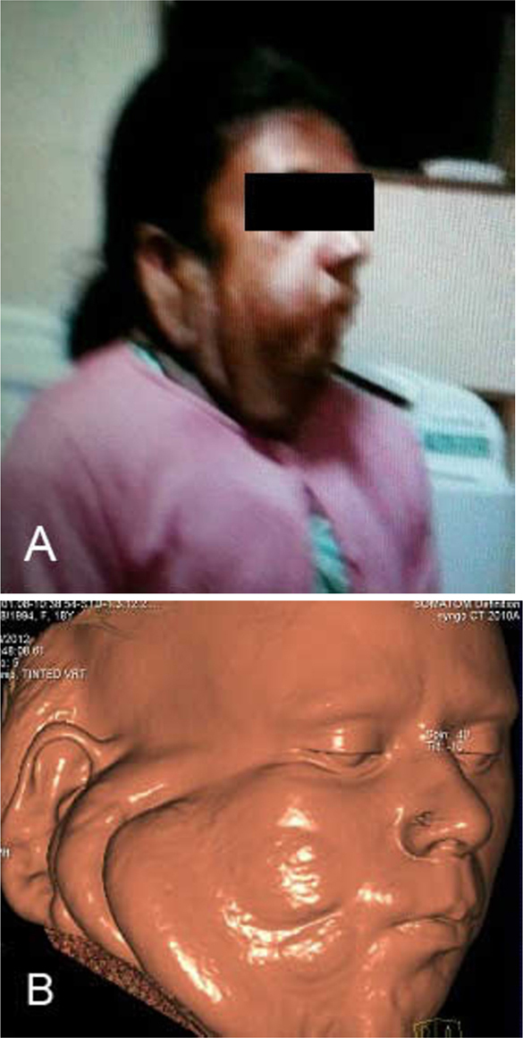
18-year-old female with plexiform neurofibroma. A. Photograph and B three-dimensional, volume-reformatted CT image of the face, showing swelling on the right side.
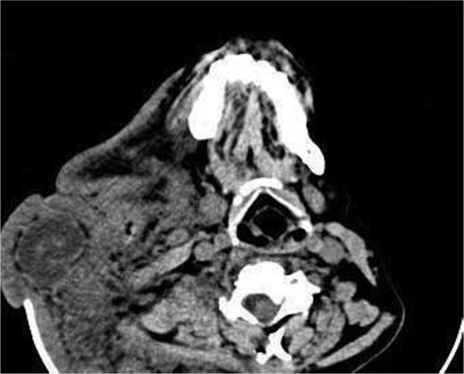
18-year-old female with plexiform neurofibroma. Axial CT image shows dysplastic changes and deformity involving right side of maxilla, mandible, and the upper cervical vertebrae.
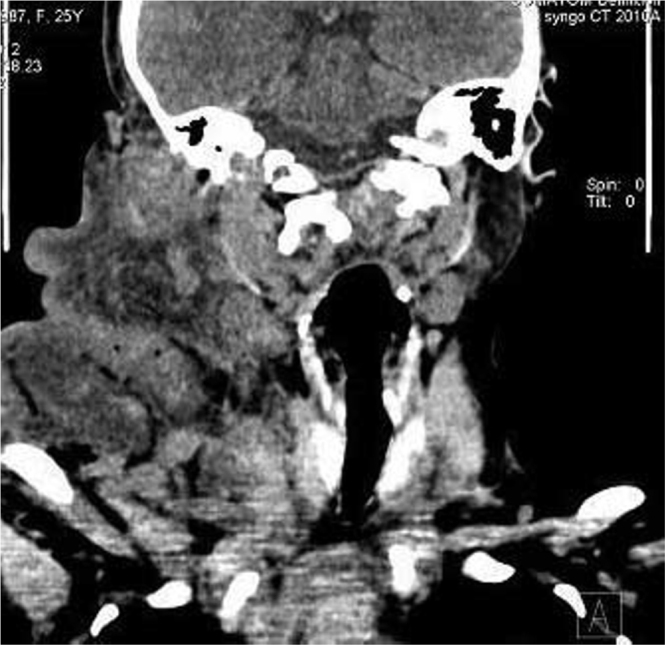
18-year-old female with plexiform neurofibroma. Coronally reformatted CT scan image extension of lesion posteriorly to the back of neck and up to the right paravertebral region.
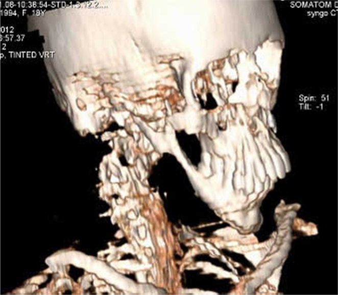
18-year-old female with plexiform neurofibroma. Three-dimensional, volume-reformatted CT image of the skull, cervical spine, and upper chest demonstrating a dysplastic right mandible.
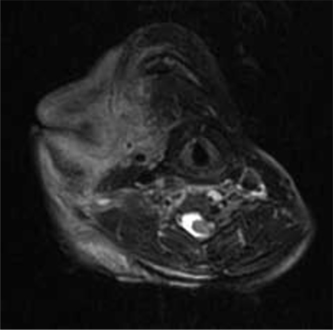
18-year-old female with plexiform neurofibroma. T2-weighted axial MRI showing overgrowth of the skin and subcutaneous tissue of the right side of the face and neck with encasement of muscle, soft tissue, and superficial blood vessels.
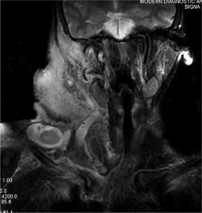
18-year-old female with plexiform neurofibroma. T2-weighted coronal MRI showing extension of the lesion up to the right paravertebral region. Note the cystic lesion measuring 30 x15 mm in the base of the right neck.
References
-
- Riccardi VM. Neurofibromatosis: Phenotype, natural history and pathogenesis. 2nd edition. Johns Hopkins University Press; Baltimore: 1992. [PubMed]
Publication types
LinkOut - more resources
Full Text Sources
Research Materials
