Case Reports
. 2015 Dec 7;7(3):714.
doi: 10.2484/rcr.v7i3.714. eCollection 2012.

Needle embolism in intravenous drug abuse

Case Reports

Needle embolism in intravenous drug abuse

Eric J Monroe et al. Radiol Case Rep. .

Abstract

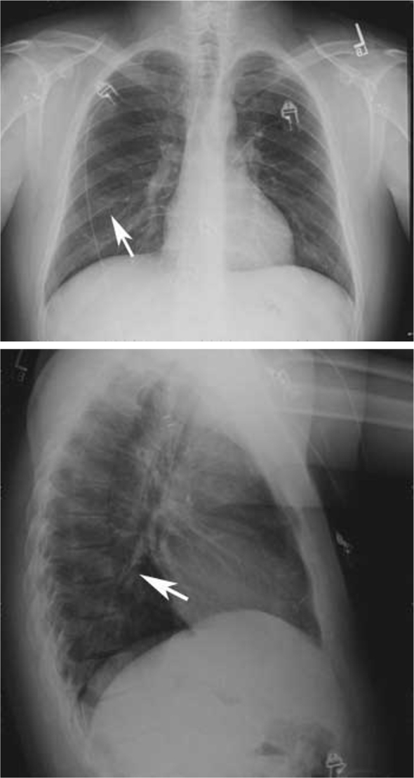
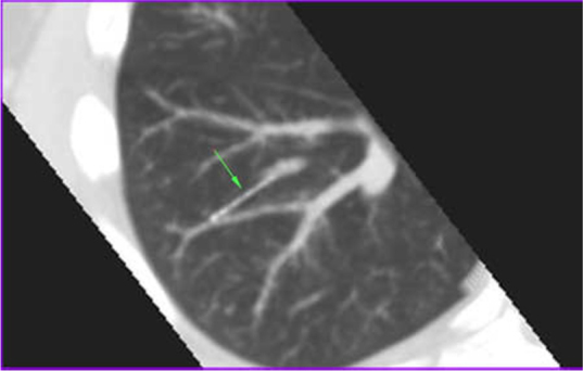
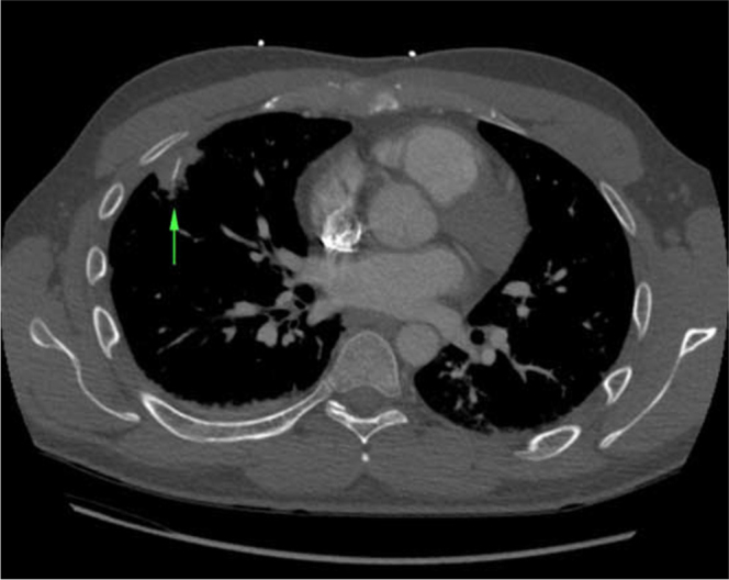
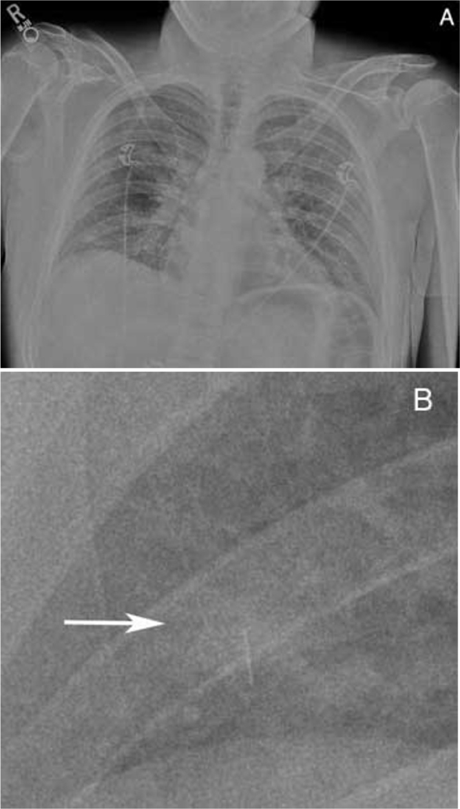
Although intravenous drug users report the breaking of a needle as a relatively common occurrence, central embolism of needle fragments occurs infrequently in the literature. Central needle embolism also poses a conundrum for the radiologist, as the needle may be easily overlooked when the clinical history is nonspecific. We present two cases of needle embolism to the lung, one complicated by inflammatory mass and progressive pleuritic chest pain requiring wedge resection. We hope that our experiences may increase radiologists' and emergency physicians' familiarity with this unusual cause of chest pain. The radiological findings are subtle and may be easily overlooked, particularly without thorough clinical history.

Keywords: CT, computed tomography; IVDU, intravenous drug use.


Figures

Figure 1
Figure 1
28-year-old man with needle embolism. Frontal and lateral radiographs of the chest demonstrate a thin linear radiodensity in the right midlung.
Figure 2
Figure 2
28-year-old man with needle embolism. Multiplanar reconstruction CT of the chest in lung windows (WL/WW -500/1400) shows the thin linear radiodensity extending from a segmental pulmonary artery into peripheral lung parenchyma (arrow).
Figure 3
Figure 3
28-year-old man with needle embolism. Axial image from pulmonary embolism protocol CT of the chest in bone windows (WL/WW 300/1500) demonstrates a linear radiodensity within an irregular consolidation in the peripheral right middle lobe (arrow).
Figure 4
Figure 4
28-year-old man with needle embolism. Frontal radiograph of the chest, A, Full field of view. B. 10X enlargement of right lung region of interest reveals a linear radiodensity in the right mid-lung (arrow) with faint surrounding consolidation.

References

    1. Substance Abuse and Mental Health Services Administration, Center for Behavioral Health Statistics and Quality. Drug abuse warning network: Detailed tables: National estimates, Drug-related emergency department visits for 2004–2009. Rockville, MD, December 28, 2010. Available at: http://www.samhsa.gov/
    1. Degenhardt L, Hall W. Extent of illicit drug use and dependence, and their contribution to the global burden of disease. Lancet. 2012;379(9810):55–70. [PubMed] - PubMed
    1. Norfolk GA, Gray SF. Intravenous drug users and broken needles–a hidden risk? Addiction. 2003;98(8):1163–1166. [PubMed] - PubMed
    1. Lewis TD, Henry DA. Needle embolus: a unique complication of intravenous drug abuse. Ann Emerg Med. 1985;14(9):906–908. [PubMed] - PubMed
    1. Angelos MG, Sheets CA, Zych PR. Needle emboli to lung following intravenous drug abuse. J Emerg Med. 1986;4(5):391–396. [PubMed] - PubMed

Publication types

LinkOut - more resources