

Multimodal Imaging in Wagner Syndrome
- PMID: 27327288
- PMCID: PMC5530878
- DOI: 10.3928/23258160-20160601-10
Multimodal Imaging in Wagner Syndrome
Abstract
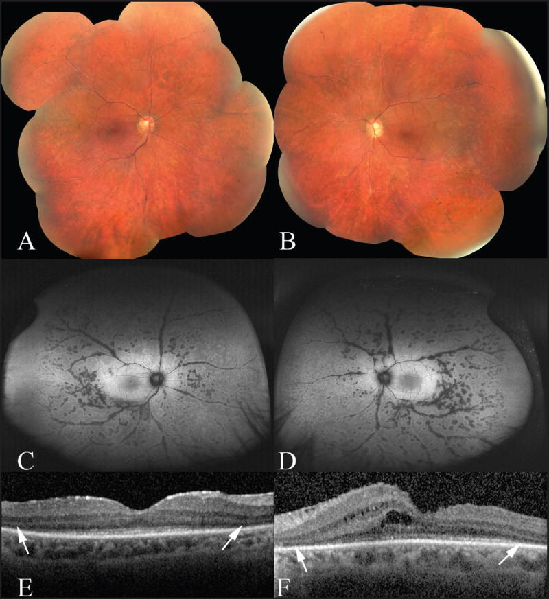
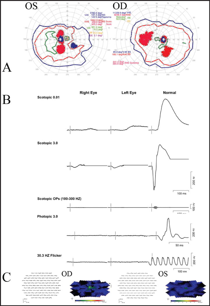
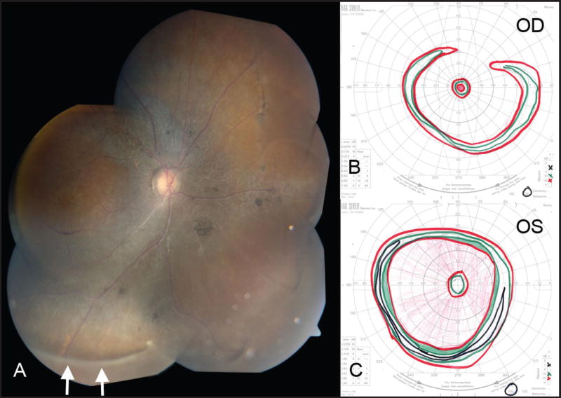
Wagner syndrome is a rare vitreoretinopathy described in a limited number of families. Here the authors describe four cases of suspected Wagner syndrome. All four cases had depressed rod and cone function on electroretinography, outer retinal disruption on spectral-domain optical coherence tomography, and constricted central visual fields with smaller isopter testing. Fundus autofluorescence performed in one patient highlighted a perivascular pattern to chorioretinal atrophy. Two patients had a history of uveitis with active cystoid macular edema. The diagnosis of Wagner syndrome was supported in three cases with genetic testing for VCAN mutations, whereas the other case harbored a variation of unknown significance in VCAN that may have been nonpathogenic. [Ophthalmic Surg Lasers Imaging Retina. 2016;47:574-579.].
Copyright 2016, SLACK Incorporated.
Figures

References
-
- Wagner H. Ein bisher unbekanntes des auges (degeneration hyaloideoretinalis hereditaria), beobachtet im Kanton Zurich. Klin Monatsbl Augenheilkd. 1938;100:840–857.
-
- Manning LM. Wagner’s hereditary vitreoretinal degeneration. Aust J Ophthalmol. 1980;8(1):29–33. - PubMed
-
- Hirose T, Lee KY, Schepens CL. Wagner’s hereditary vitreoretinal degeneration and retinal detachment. Arch Ophthalmol. 1973;89(3):176–185. - PubMed
-
- Graemiger RA, Niemeyer G, Schneeberger SA, Messmer EP. Wagner vitreoretinal degeneration. Follow-up of the original pedigree. Ophthalmology. 1995;102(12):1830–1839. - PubMed
-
- Brown DM, Graemiger RA, Hergersberg M, et al. Genetic linkage of Wagner disease and erosive vitreoretinopathy to chromosome 5q13–4. Arch Ophthalmol. 1995;113(5):671–675. - PubMed
Publication types
MeSH terms
Substances
Supplementary concepts
Grants and funding
LinkOut - more resources
Full Text Sources
Other Literature Sources
