. 2016 Aug:149:75-83.
doi: 10.1016/j.exer.2016.06.010. Epub 2016 Jun 18.

Whole-eye electrical stimulation therapy preserves visual function and structure in P23H-1 rats

Affiliations

Whole-eye electrical stimulation therapy preserves visual function and structure in P23H-1 rats

Adam M Hanif et al. Exp Eye Res. 2016 Aug.

Abstract

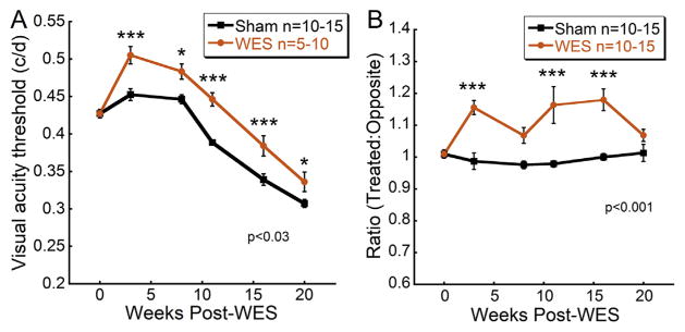
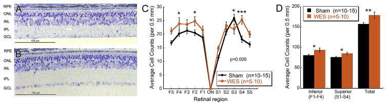
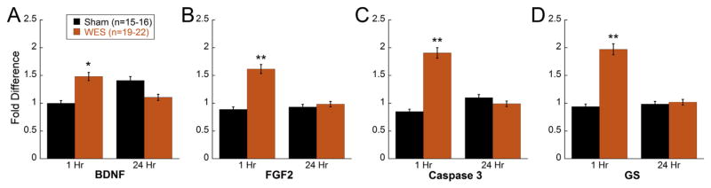
Low-level electrical stimulation to the eye has been shown to be neuroprotective against retinal degeneration in both human and animal subjects, using approaches such as subretinal implants and transcorneal electrical stimulation. In this study, we investigated the benefits of whole-eye electrical stimulation (WES) in a rodent model of retinitis pigmentosa. Transgenic rats with a P23H-1 rhodopsin mutation were treated with 30 min of low-level electrical stimulation (4 μA at 5 Hz; n = 10) or sham stimulation (Sham group; n = 15), twice per week, from 4 to 24 weeks of age. Retinal and visual functions were assessed every 4 weeks using electroretinography and optokinetic tracking, respectively. At the final time point, eyes were enucleated and processed for histology. Separate cohorts were stimulated once for 30 min, and retinal tissue harvested at 1 h and 24 h post-stimulation for real-time PCR detection of growth factors and inflammatory and apoptotic markers. At all time-points after treatment, WES-treated rat eyes exhibited significantly higher spatial frequency thresholds than untreated eyes. Inner retinal function, as measured by ERG oscillatory potentials (OPs), showed significantly improved OP amplitudes at 8 and 12 weeks post-WES compared to Sham eyes. Additionally, while photoreceptor segment and nuclei thicknesses in P23H-1 rats did not change between treatment groups, WES-treated eyes had significantly greater numbers of retinal ganglion cell nuclei than Sham eyes at 20 weeks post-WES. Gene expression levels of brain-derived neurotrophic factor (BDNF), caspase 3, fibroblast growth factor 2 (FGF2), and glutamine synthetase (GS) were significantly higher at 1 h, but not 24 h after WES treatment. Our findings suggest that WES has a beneficial effect on visual function in a rat model of retinal degeneration and that post-receptoral neurons may be particularly responsive to electrical stimulation therapy.

Keywords: Electrical stimulation therapy; Neuroprotection; Retinal degeneration; Retinitis pigmentosa.


Figures

Fig. 1
Fig. 1. Electrostatic model used for finite element analysis of the WES electrode configuration
A: Cross-sectional view, showing meshed model, white circles indicating electrode positions on cornea and in mouth. B: Contour plot of voltage; white squares mark measurement sites used in model validation (color scale 0–5.52e−3 V). C: Isolated photoreceptor layer from the electrostatic model plotting current density (color scale 0.396e−5–0.142e−3 A/m2).
Fig. 2
Fig. 2. WES preserves visual acuity in treated eyes
Visual acuity was preserved in WES-treated eyes. (A) Visual acuity in WES (n = 5–10) and Sham (n = 10–15) eyes was measured with OKT. At all time points after baseline, WES-treated eyes exhibited significantly greater preservation of spatial frequency thresholds (Two way repeated measures ANOVA, F(5,129) = 2.67; p = 0.027). (B) Spatial frequency thresholds of each treated eye were divided by threshold values measured from their contralateral, untreated eyes. Eyes that received WES had significant increased visual acuity threshold ratios compared to contralateral eyes (Two way repeated measures ANOVA, F(5,107) = 7.744; p < 0.001). Post-hoc comparisons: *p < 0.05, **p < 0.01, ***p < 0.001.
Fig. 3
Fig. 3. WES preserves inner retinal function
(A) Representative dark-adapted OP responses to a bright flash (2.9 log cd s/m2) across time in treated eyes for both WES and Sham groups. Differences in peak amplitudes between experimental groups were perceivable at 8 and 12 weeks post-WES. (B) OP2 amplitudes were significantly higher in WES-treated eyes in response to the brighter flash stimuli at 8 weeks post-WES (Two way repeated measures ANOVA, F(11,107) = 2.318; p = 0.016) (C) Additionally, OP2 amplitudes remained significantly higher in WES-treated eyes compared to Sham for the brighter stimuli at 12 weeks post-WES (Two way repeated measures ANOVA, F(11,155) = 2.428; p = 0.009) (D) Maximum OP2 amplitudes elicited from the brightest flash stimuli showed a trend for increased amplitudes in the WES-treated rats at 8 and 12 weeks compared to Sham, but no statistically significant differences were found. Post-hoc comparisons, *p < 0.05, **p < 0.01, ***p < 0.001.
Fig. 4
Fig. 4. WES preserves RGCs
Representative images of retinal cross sections from Sham (A) and WES (B) treated eyes. Cellular density in the RGC layer is visibly more robust in WES treated eyes, indicating that electrical stimulation of the whole eye preserved retinal morphology in the P23H-1 model. (C) WES-treated eyes yielded greater RGC counts than Sham treated eyes (Two way repeated measures ANOVA, F(9,551) = 2.638; p = 0.005) (D) Sums of inferior (Student’s t-test, p = 0.013) and superior (Student’s t-test; p = 0.027) regions 1–4 between groups also revealed significantly greater nuclei in the RGC layer of WES treated eyes. Sums of all regions (Total) showed more RGC layer cells in the WES-treated eyes (Student’s t-test; p = 0.005). Post-hoc comparisons: *p < 0.05, **p < 0.01, ***p < 0.001.
Fig. 5
Fig. 5. WES upregulates specific growth factors and apoptotic markers
RT-PCR analysis revealed elevated expression of (A) Bdnf (Two way ANOVA, F(1,71) = 7.064; p = 0.01), (B) Fgf2 (Two way ANOVA, F(1,65) = 4.956; p = 0.03), (C) Casp3 (Two way ANOVA, F(1,69) = 5.223; p = 0.026), and (D) Gs (Two way ANOVA, F(1,66) = 5.197; p = 0.03) in P23H-1 rats (aged 4 weeks) subjected to one 30-min session of WES and sacrificed 1 h after treatment compared to Sham eyes. No significant differences in gene expression were detected in retinas harvested 24 h after WES therapy. Asterisks indicate post-hoc comparisons: *p < 0.05, **p < 0.01.

References

    1. Allen RS, Sayeed I, Cale HA, Morrison KC, Boatright JH, Pardue MT, Stein DG. Severity of middle cerebral artery occlusion determines retinal deficits in rats. Exp Neurol. 2014;254:206–215. - PMC - PubMed
    1. Andreucetti D, Rossi R, Petrucci C. An Internet Resource for the Calculation of the Dielectric Properties of Body Tissues in the Frequency Range 10 Hz – 100 GHz. IFAC-CNR; Florence, Italy: 1997. http://niremf.ifac.cnr.it/tissprop/
    1. Barnett NL, Pow DV, Robinson SR. Inhibition of Muller cell glutamine synthetase rapidly impairs the retinal response to light. Glia. 2000;30:64–73. - PubMed
    1. Berson EL, Rosner B, Sandberg MA, Dryja TP. Ocular findings in patients with autosomal dominant retinitis pigmentosa and a rhodopsin gene defect (Pro-23-His) Arch Ophthalmol. 1991;109:92–101. - PubMed
    1. Burroughs SL, Kaja S, Koulen P. Quantification of deficits in spatial visual function of mouse models for glaucoma. Investig Ophthalmol Vis Sci. 2011;52:3654–3659. - PMC - PubMed

Publication types

MeSH terms

Substances