. 2016 Jun 21;12(6):e1005700.
doi: 10.1371/journal.ppat.1005700. eCollection 2016 Jun.

KIF5B and Nup358 Cooperatively Mediate the Nuclear Import of HIV-1 during Infection

Affiliations

KIF5B and Nup358 Cooperatively Mediate the Nuclear Import of HIV-1 during Infection

Adarsh Dharan et al. PLoS Pathog. .

Abstract

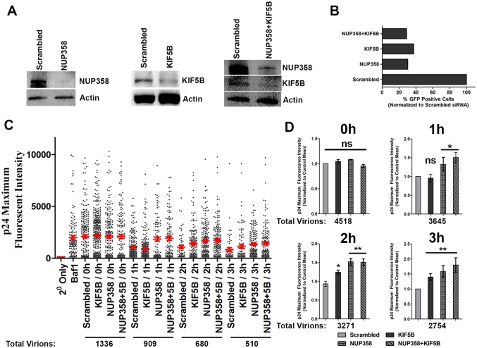
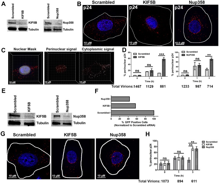
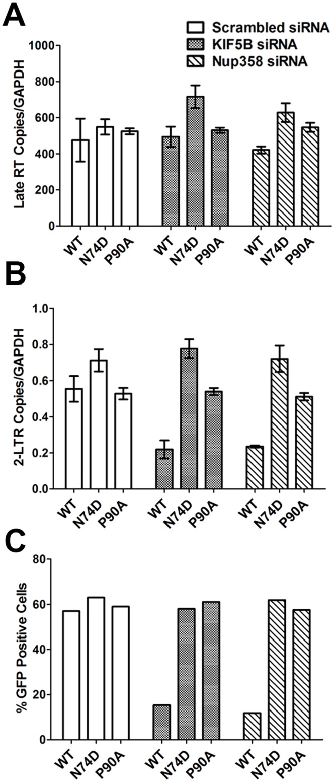
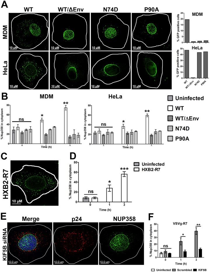
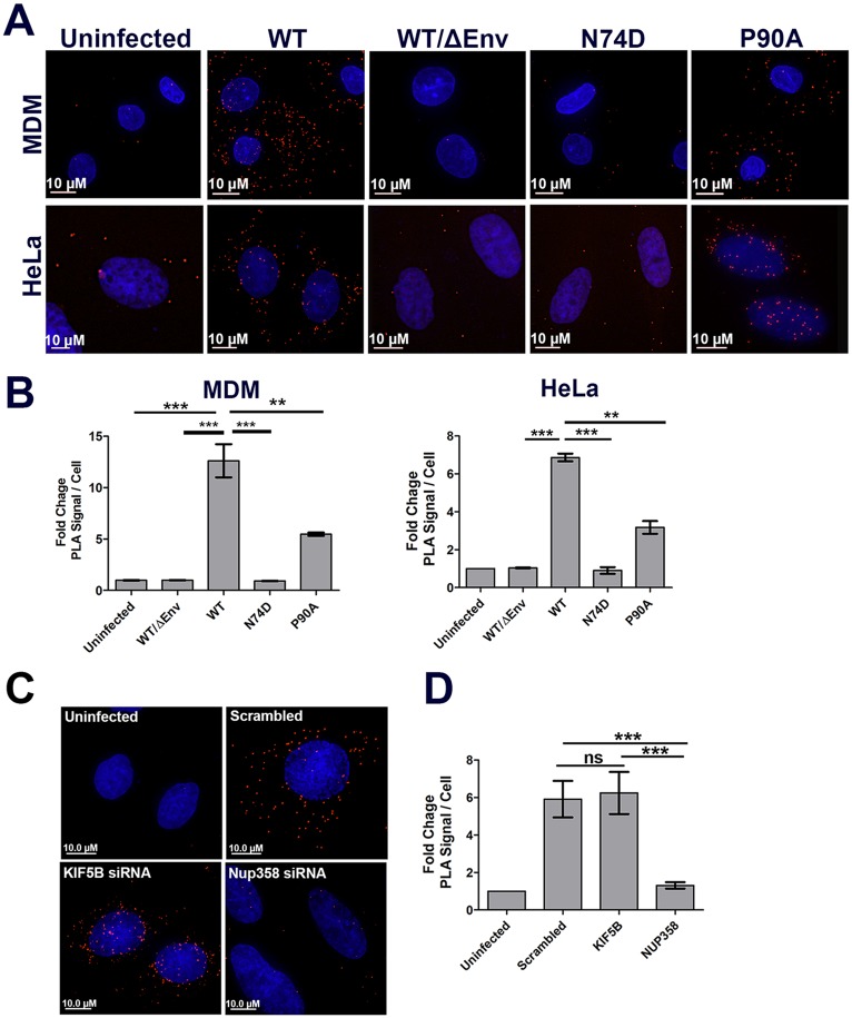
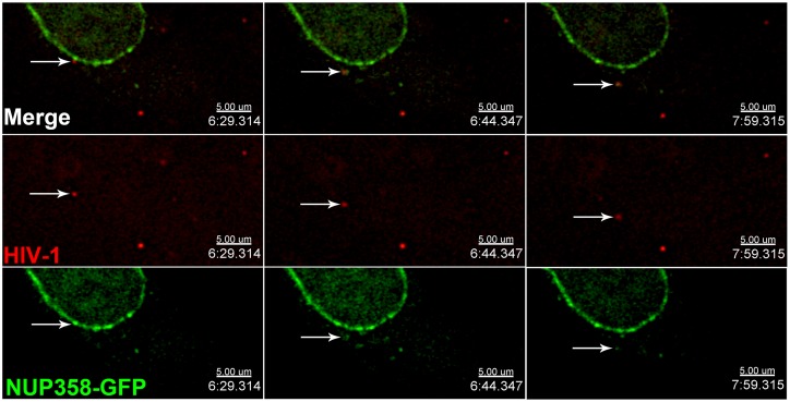
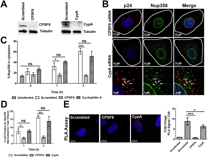
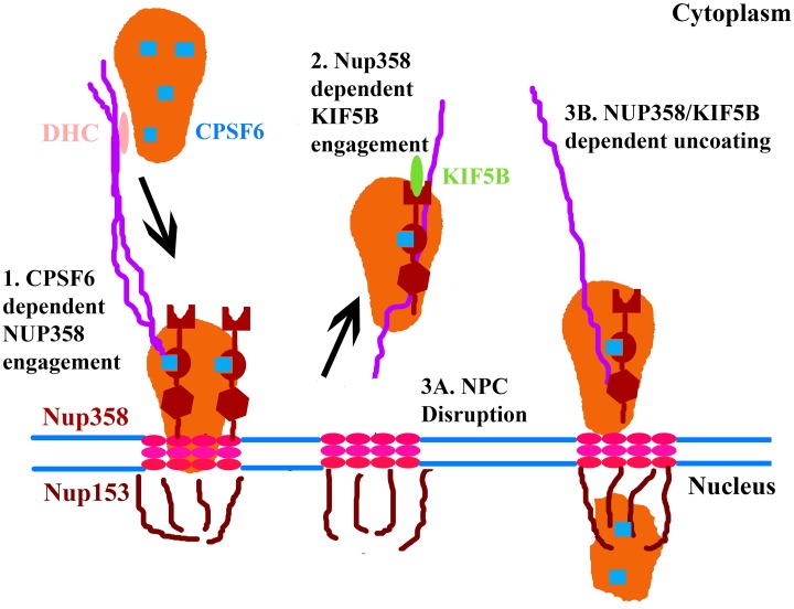
Following envelope mediated fusion, the HIV-1 core is released into the cytoplasm of the target cell and undergoes a series of trafficking and replicative steps that result in the nuclear import of the viral genome, which ultimately leads to the integration of the proviral DNA into the host cell genome. Previous studies have found that disruption of microtubules, or depletion of dynein or kinesin motors, perturb the normal uncoating and trafficking of the viral genome. Here, we show that the Kinesin-1 motor, KIF5B, induces a relocalization of the nuclear pore component Nup358 into the cytoplasm during HIV-1 infection. This relocalization of NUP358 is dependent on HIV-1 capsid, and NUP358 directly associates with viral cores following cytoplasmic translocation. This interaction between NUP358 and the HIV-1 core is dependent on multiple capsid binding surfaces, as this association is not observed following infection with capsid mutants in which a conserved hydrophobic binding pocket (N74D) or the cyclophilin A binding loop (P90A) is disrupted. KIF5B knockdown also prevents the nuclear entry and infection by HIV-1, but does not exert a similar effect on the N74D or P90A capsid mutants which do not rely on Nup358 for nuclear import. Finally, we observe that the relocalization of Nup358 in response to CA is dependent on cleavage protein and polyadenylation factor 6 (CPSF6), but independent of cyclophilin A. Collectively, these observations identify a previously unappreciated role for KIF5B in mediating the Nup358 dependent nuclear import of the viral genome during infection.


Conflict of interest statement

The authors have declared that no competing interests exist.

Figures

Fig 1
Fig 1. KIF5B and NUP358 facilitate HIV-1 uncoating.
(A) HeLa cells were transfected with siRNA’s targeting Nup358, KIF5B or both (Nup358+KIF5B). Expression of the indicated proteins were determined by western blot 96h post transfection.(B) siRNA treated HeLa cells infected with VSVg-HIV-1 GFP (MOI 0.8) and infectivity measured by determining the percent of GFP positive cells by FACS. (C) HeLa cells treated with scrambled, Nup358, KIF5B or Nup358+5B siRNA for 96h were synchronously infected with S15-mCherry/GFP-Vpr VSVg-HIV-1. Cells were fixed at the indicated time point, and the p24 intensities associated with individual virions lacking the S15 membrane label (1–3 hours PI) or all virions (controls and 0 hr points) are shown. Bafilomycin A1 (Baf1), inhibits VSVg mediated fusion and was used as a control in these experiments. Red line is the average p24 intensity measured for all fused viruses at the indicated time point. At least 20 cells were imaged at each time point. Error bars represent SEM.(D)Data from three independent experiments, as shown in C, were normalized to the mean p24 intensity observed in control siRNA transfected cell and averaged. **p<0.01, *p<0.05, ns = not significant. Data is representative of three or more independent experiments.
Fig 2
Fig 2. KIF5Band Nup358 depletion lead to the perinuclear accumulation of HIV-1 cores.
HeLa cells were transfected with siRNA’s targeting Nup358, KIF5B or a scrambled siRNA sequence.(A) Western blot for KIF5B or NUP358 96h following siRNA transfection (B)siRNA depleted cells were synchronously infected withVSVg-R7ΔEnvGFP(MOI 0.6). Cells were fixed 0, 1and 3h following a synchronized infection and stained for HIV-1 capsid protein p24 (red) and DAPI (blue) for cell nuclei. A representative image at 3h post infection is depicted.(C)Quantification process employed to detect perinuclear and cytoplasmic p24 protein levels. Nuclear mask generated based on the DAPI channel (left image) and perinuclear signal quantified by masking all signal inside (middle image) or outside right image) of the nuclear mask. (D)Percentage ofp24 puncta in the perinuclear region in KIF5B and Nup358 depleted cells, quantified as described in (C). TZM-bl cells were transfected with siRNA’s targeting Nup358, KIF5B or a scrambled siRNA sequence.(E) Western blot for KIF5B or NUP358 72h following siRNA transfection. siRNA depleted cells were synchronously infected with HXB2-R7ΔEnvGFP(MOI 0.32). (F)Infectivity was assessed 48 hours following infection. (G,H) Cells were fixed 0, 1and 3h following a synchronized infection and stained for HIV-1 capsid protein p24 (red) and DAPI (blue) for cell nuclei. A representative image at 3h post infection is depicted. (H) Percentage ofp24 puncta in the perinuclear region in KIF5B and Nup358 depleted cells, quantified as described in (C). 20 or more cells was analyzed at each time point. Error bars represent the SEM of three experiments. The total number of virions analyzed at each time point is shown below each graph. ***p<0.001, *p<0.05, ns = not significant. Data is representative of three or more independent experiments.
Fig 3
Fig 3. HIV-1 capsid influences the dependence on KIF5B and Nup358 mediated nuclear import during infection.
HeLa cells treated with scrambled, Nup358 or KIF5B siRNA for 72h were subjected to synchronized infected with 100ng of VSVg pseudotyped HIV-1 reporter virus bearing either the wildtype (WT) CA or N74D and P90A CA mutants. Cells were collected 24 post infection and real time PCR performed using specific primers to quantify Late RT (A) and 2-LTR circle (B). Error bars represent the standard deviation of single samples run in triplicate. (C) GFP expression 48h post infection, as measured by flow cytometry of 10,000 cells. The data shown here is representative of three independent experiments.
Fig 4
Fig 4. HIV-1 infection induces Nup358 relocalization.
(A,B) Monocyte derived macrophages (MDM) and HeLa cells were synchronously infected with VSVg pseudotyped HIV-1 reporter virus (MOI 0.3 for MDM and MOI 0.6 for HeLa cells) bearing either the wildtype (WT) CA or N74D and P90A CA mutants. Cells were fixed at 0, 1 or 3h (shown) post infection and stained for Nup358 (green). Infection for each cell type is shown (B)The fraction of Nup358 signal in the cytoplasm at the indicated time PI, measured as in 2C.(C,D)TZM-Bl cells were synchronously infected with R7ΔEnvGFPpseudotyped with the HXB2 envelope protein (MOI 0.32). Cells were fixed 0, 1 and 3h (shown) post infection and stained for Nup358. (D) The fraction of Nup358 signal in the cytoplasm at the indicated time PI.(E,F)HeLa cells were transfected with KIF5B specific or scrambled control siRNA and synchronously infected with R7ΔEnvGFPpseudotyped with VSV-g (MOI 0.6)96 hours following siRNA transfection. Cells were fixed 0, 1 and 3h (shown) post infection and stained for Nup358.(F)The fraction of Nup358 signal in the cytoplasm at the indicated time PI.20 or more cells were analyzed for each sample. Error bars represent the SEM of three independent experiments. (**p<0.01, *p<0.05, ns = not significant). Data is representative of three or more independent experiments.
Fig 5
Fig 5. Nup358 and HIV-1 cores colocalize in the cytoplasm during infection.
(A)MDMs were synchronously infected with HIV-1 GFP pseudotyped with VSV-g bearing either the wildtype (WT) CA or N74D and P90A CA mutants (MOI 0.3).Cells fixed 1 and 3h post infection and stained for viral capsid protein p24 (red) and Nup358 (green). Depicted a representative image at 3h post infection of WT virus (left panel) and an enlarged view of the same image showing colocalization of Nup358 and p24 (right panel and indicated by arrows). (B) Similar experiments as A performed on HeLa cells (MOI 0.6). (C,D) Quantification of the percent p24 colocalizing with Nup358 in MDM cells (C) or HeLa cells (D).20 or more cells were analyzed in each sample. Error bars represent the SEM of three independent experiments. (**p<0.01, *p<0.05, ns = not significant). Data is representative of three or more independent experiments.
Fig 6
Fig 6. Nup358 associates with HIV-1 cores in the cytoplasm during infection.
(A) MDM or HeLa cells were synchronously infected HIV-1 GFP pseudotyped with VSV-g bearing either the wildtype (WT) CA or N74D and P90A CA mutants(MDMs MOI 0.3, HeLa MOI 0.6).Cells fixed at 3h post infection, incubated with primary antibodies to Nup358 and HIV-1 CA, followed by secondary antibodies specific for these antibodies conjugated to the PLUS and MINUS PLA oligonucleotides. Each red fluorescent puncta represents a positive PLA signal generated by the interaction of PLUS and MINUS oligonucleotides bound to secondary antibodies. (B) Quantification of PLA signal in MDMs and HeLa cells, as measured by the average fold increase in PLA signal, relative to uninfected control, in three independent experiments.(C) HeLa cells were transfected with siRNA’s targeting KIF5B, Nup358 or scrambled control siRNA and synchronously infected with R7ΔEnvGFPpseudotyped with VSV-g (MOI 0.6) 96 hours following siRNA transfection. Cells fixed at 3h post infection and PLA assay performed as described in (A). (D) Quantification of PLA signal in the siRNA knockdown cells as in (B). 20 or more cells were analyzed in each experiment. Error bars represent the SEM(**p<0.01, *p<0.05, ns = not significant). Data is representative of three or more independent experiments.
Fig 7
Fig 7. Anterograde trafficking of HIV-1 cores during infection.
HeLa cells transfected with a NUP358-GFP expressing BAF were synchronously infected with GIR labelled HIV-1 viral particles (MOI 0.3). 2 hours after infection, cells were imaged every 15 seconds for 10 minutes. Shown are individual z-planes showing a virus observed to traffic away from the nucleus during the acquisition period while associated with NUP358-GFP.
Fig 8
Fig 8. Nup358 association with HIV-1 cores is CPSF6 dependent and Cyclophilin A independent.
HeLa cells transfected with scrambled siRNA or siRNA specific for CPSF6 or CypA.(A)Western blot for CPSF6 or CypA 96h following siRNA transfection. (B)siRNA depleted cells were synchronously infected with VSVg-R7ΔEnvGFP(MOI 0.6). Cells were fixed 0, 1 and 3h post infection stained for HIV-1 capsid protein p24 (red) and NUP358 (green).Depicted a representative image at 3h post infection. Nup358 and CA colocalization following CypA depletion shown in bottom panel. (C)The fraction of Nup358 signal in the cytoplasm at the indicated time PI. (D) Quantification of CA and Nup358 signal colocalization in the siRNA depleted cells. (E) PLA of Nup358 and CA, performed on the siRNA depleted cells fixed 3 hours following synchronous infection, and quantification of average fold increase in PLA signal. 20 or more cells were analyzed in each experiment. Error bars represent the SEM (**p<0.01, *p<0.05, ns = not significant). Data is representative of three or more independent experiments.
Fig 9
Fig 9. Model of KIF5B mediated nuclear import of HIV-1.
(1) During the early steps of infection, CPSF6 binding to the viral core occurs. This interaction between CPSF6 (blue squares) and the viral core and cytoplasmic dynein heavy chain (DHC) mediated trafficking along microtubules is necessary to facilitate an interaction between NUP358 and the core. (2)Core engagement by Nup358 is necessary to trigger the KIF5B mediated trafficking of both the core and Nup358 away from the nuclear pore. There are two potential mechanisms by which this KIF5B trafficking facilitates the nuclear import of the viral genome. (3A)First, NPC disruption, induced through KIF5B mediated dislocation of Nup358 away from the viral core, may facilitate the ability of the PIC to enter the nucleus by enhancing nuclear permeability, as previously demonstrated in the case of adenovirus infection [52]. (3B)Second, cytoplasmic uncoating, cooperatively mediated by NUP358 and KIF5B in the cytoplasm, may ultimately reduce the size of the PIC to dimensions compatible with translocation across the nuclear pore.

References

    1. Yamashita M, Emerman M (2004) Capsid is a dominant determinant of retrovirus infectivity in nondividing cells. J Virol 78: 5670–5678. - PMC - PubMed
    1. Yamashita M, Perez O, Hope TJ, Emerman M (2007) Evidence for direct involvement of the capsid protein in HIV infection of nondividing cells. PLoS Pathog 3: 1502–1510. - PMC - PubMed
    1. Campbell EM, Hope TJ (2015) HIV-1 capsid: the multifaceted key player in HIV-1 infection. Nat Rev Microbiol 13: 471–483. 10.1038/nrmicro3503 - DOI - PMC - PubMed
    1. Yan N, Regalado-Magdos AD, Stiggelbout B, Lee-Kirsch MA, Lieberman J (2010) The cytosolic exonuclease TREX1 inhibits the innate immune response to human immunodeficiency virus type 1. Nat Immunol 11: 1005–1013. 10.1038/ni.1941 - DOI - PMC - PubMed
    1. Rasaiyaah J, Tan CP, Fletcher AJ, Price AJ, Blondeau C, et al. (2013) HIV-1 evades innate immune recognition through specific cofactor recruitment. Nature 503: 402–405. 10.1038/nature12769 - DOI - PMC - PubMed