Review
. 2017 Oct;105(7):2162-2173.
doi: 10.1002/jbm.b.33734. Epub 2016 Jun 21.

The biological response to orthopaedic implants for joint replacement: Part I: Metals

Affiliations
Review

The biological response to orthopaedic implants for joint replacement: Part I: Metals

Emmanuel Gibon et al. J Biomed Mater Res B Appl Biomater. 2017 Oct.

Abstract

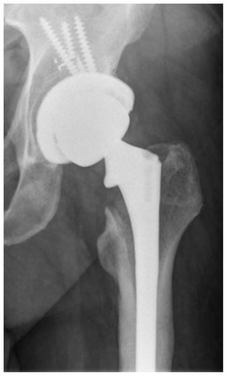
Joint replacement is a commonly performed, highly successful orthopaedic procedure, for which surgeons have a large choice of different materials and implant designs. The materials used for joint replacement must be both biologically acceptable to minimize adverse local tissue reactions, and robust enough to support weight bearing during common activities of daily living. Modern joint replacements are made from metals and their alloys, polymers, ceramics, and composites. This review focuses on the biological response to the different biomaterials used for joint replacement. In general, modern materials for joint replacement are well tolerated by the body as long as they are in bulk (rather than in particulate or ionic) form, are mechanically stable and noninfected. If the latter conditions are not met, the prosthesis will be associated with an acute/chronic inflammatory reaction, peri-prosthetic osteolysis, loosening and failure. This article (Part 1 of 2) is dedicated to the use of metallic devices in orthopaedic surgery including the associated biological response to metallic byproducts is a review of the basic science literature regarding this topic. © 2016 Wiley Periodicals, Inc. J Biomed Mater Res Part B: Appl Biomater, 105B: 2162-2173, 2017.

Keywords: biological response; biomaterials; foreign body response; inflammation; orthopaedic implants.


Figures

FIGURE 1
FIGURE 1
The passivation layer is an oxidized metal (black). The passivation layer prevents further degradation of the underlying coalloy (Implant, gray). Metal implanted into the body has reacted with oxygen and is passivated. However, damage to the passivation layer results in exposure of the reactive coalloy surface, which reacts oxygen (O2) and water (H2O) resulting in repassivation. Illustration created by Chrisoula Toupadakis Skouritakis, Ph.D.
FIGURE 2
FIGURE 2
Examples of the types of modular junctions in total hip arthroplasty: (A) S-ROM illustrating proximal metaphyseal modularity, (B) dual modular design illustrating neck–stem modularity, (C) fluted tapered modular stem illustrating distal diaphyseal modularity, and (D) total femoral replacement illustrating modularity at its extreme. Note, examples A–D all illustrate head–neck modularity using the trunnion of the stem to accept a modular femoral head.
FIGURE 3
FIGURE 3
The junction between the passivated femoral head and stem trunnion (dark gray) results in the microisolation of the corrosive environment that establishes an oxygen gradient between the interstitial fluid (“normoxic”) and the crevice fluid (“anoxic”). (A) The lack of oxygen inside the crevice leads to the separation of the anodic process (metal dissolution inside the crevice) and the cathodic process (oxygen reduction outside the crevice). The excess of positive ions in the crevice solution is balanced by an influx of negatively charged ions, primarily chloride ions (Cl). The electrons generated by the anodic process is picked up by oxygen at the cathodic sites on the metal surface outside the crevice. (B) The hydrolysis of the chromium produces chromium ion (Cr3+) during the anodic process results in hydrogen ion (H+) generation inside the crevice, dropping the pH of the microenvironment and accelerating metal ion release. (C) As the crevice solution becomes more acidic, the hydrolysis of chromium becomes less and less complete, and the rate of acidification decreases. (D) As the critical pH is reached, the chromium ions are no longer hydrolyzed and crevice corrosion progresses at the rate of metal ion (CO2+, Cr3+, and so forth) diffusion. The metal ions then can react with organic and inorganic anions in the vicinity of the crevice opening, and—in the case of chromium ions—form insoluble precipitates containing chromium oxide (Cr2O3) and chromium phosphate (CrPO4) on the metal surface. Illustration created by Chrisoula Toupadakis Skouritakis, Ph.D.
FIGURE 4
FIGURE 4
When a modular junction is loaded, the implant flexes (slip region) while remaining together (stick region). This micromotion can physically disrupt the passivation layer, leading to metal wear or implant fracture. Micromotion does not equate to loosening. Illustration created by Chrisoula Toupadakis Skouritakis, Ph.D.
FIGURE 5
FIGURE 5
(A) Hip resurfacing arthroplasty and (B) total hip arthroplasty with a metal head and metal acetabular liner are types of metal-on-metal articulations in total joint arthroplasty.
FIGURE 6
FIGURE 6
A polar bearing prosthesis leads to proper bedding-in and low steady-state wear as well as fluidfilm lubrication assuming proper implant positioning. Illustration created by Chrisoula Toupadakis Skouritakis, Ph.D.
FIGURE 7
FIGURE 7
4× magnification of the diffuse perivascular infiltrates of T-and B-lymphocytes along with plasma cells as well as macrophages with or without metal debris that is characteristic of aseptic lymphocyte dominated vasculitis-associated lesions (ALVAL).
FIGURE 8
FIGURE 8
T1-weighted coronal section from a metal artifact reduction sequence (MARS) MRI demonstrating large pseudotumors involving the gluteus maximus muscle and lateral left hip.
FIGURE 9
FIGURE 9
Severe osteolysis of the calcar and failure of acetabular fixation.

References

    1. Richards RG, Moriarty TF, Miclau T, McClellan RT, Grainger DW. Advances in biomaterials and surface technologies. J Orthop Trauma. 2012;26:703–707. - PubMed
    1. Epinette JA, Manley MT. Hydroxyapatite-coated total knee replacement: Clinical experience at 10 to 15 years. J Bone Joint Surg Br. 2007;89:34–38. - PubMed
    1. Attar FG, Khaw F-M, Kirk LMG, Gregg PJ. Survivorship analysis at 15 years of cemented press-fit condylar total knee arthroplasty. J Arthroplasty. 2008;23:344–349. - PubMed
    1. Melton JTK, Mayahi R, Baxter SE, Facek M, Glezos C. Long-term outcome in an uncemented, hydroxyapatite-coated total knee replacement: A 15- to 18-year survivorship analysis. J Bone Joint Surg Br. 2012;94:1067–1070. - PubMed
    1. El Masri F, Kerboull L, Kerboull M, Courpied JP, Hamadouche M. Is the so-called ‘French paradox’ a reality? Long-term survival and migration of the Charnley–Kerboull stem cemented line-to-line. J Bone Joint Surg Br. 2010;92:342–348. - PubMed