Review
. 2016 Jul 7;8(25):12471-503.
doi: 10.1039/c5nr08691d. Epub 2016 Jun 20.

Photonanomedicine: a convergence of photodynamic therapy and nanotechnology

Affiliations
Review

Photonanomedicine: a convergence of photodynamic therapy and nanotechnology

Girgis Obaid et al. Nanoscale. .

Abstract

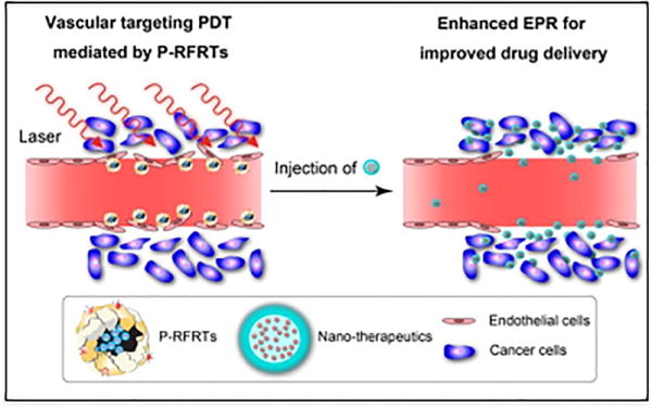
As clinical nanomedicine has emerged over the past two decades, phototherapeutic advancements using nanotechnology have also evolved and impacted disease management. Because of unique features attributable to the light activation process of molecules, photonanomedicine (PNM) holds significant promise as a personalized, image-guided therapeutic approach for cancer and non-cancer pathologies. The convergence of advanced photochemical therapies such as photodynamic therapy (PDT) and imaging modalities with sophisticated nanotechnologies is enabling the ongoing evolution of fundamental PNM formulations, such as Visudyne®, into progressive forward-looking platforms that integrate theranostics (therapeutics and diagnostics), molecular selectivity, the spatiotemporally controlled release of synergistic therapeutics, along with regulated, sustained drug dosing. Considering that the envisioned goal of these integrated platforms is proving to be realistic, this review will discuss how PNM has evolved over the years as a preclinical and clinical amalgamation of nanotechnology with PDT. The encouraging investigations that emphasize the potent synergy between photochemistry and nanotherapeutics, in addition to the growing realization of the value of these multi-faceted theranostic nanoplatforms, will assist in driving PNM formulations into mainstream oncological clinical practice as a necessary tool in the medical armamentarium.


Figures

Figure 1
Figure 1
Distribution of the total number of clinical trials using nanoparticles and liposomes in the five highest contributing regions of the world.
Figure 2
Figure 2
A) A schematic representation of the Jablonski diagram showing how PDT and fluorescence induced is by the irradiation of a PS. The PS in the ground state (S0) becomes excited by incident light (hνA or hνB) to the S1 or S2 singlet excited states. The excited PS can relax to S0 by the radiative fluorescence emission of photons (hνF), which can be used for imaging and diagnostics, or can undergo a spin forbidden process termed intersystem crossing. Through the spin flip of the excited PS, the molecule occupies a long-lived triplet excited state (T1), from which photochemical reactions occur that result in the production of cytotoxic RMS, including 1O2, that is used for PDT. B) A diagrammatic representation of the use of a PS for PDT and imaging techniques. Through type I and type II photochemical reactions, the excited sensitizer generates cytotoxic 1O2 and RMS from ground state triplet oxygen (3O2) and various biochemical substrates. The concomitant fluorescence of the PSs allows for imaging and diagnostic uses, emphasizing their inherent theranostic capacity.
Figure 3
Figure 3
A representation of the steps taken during a clinical PDT procedure using various PS formulations and the structural and functional information obtained using optical imaging techniques that enables treatment prediction and guidance. Following intravenous administration of the PS, an appropriate PS-light interval is required prior to irradiation using localized NIR light delivery. Through spatially confined PDT action on the tumor, the disease tissue is destroyed. Figure adapted with permission from Mallidi et al. Optical Imaging, Photodynamic Therapy and Optically Triggered Combination Treatments, The Cancer Journal, 21 (3), p194-205. (Copyright © 2015 Wolters Kluwer Health, Inc.)
Figure 4
Figure 4
A schematic diagram of the visionary theranostic nanoconstruct that combines a fluorescence-based theranostic or imaging agent and a therapeutic drug encapsulated within. The surface is grafted with a targeting ligand to enable the molecular selectivity of the theranostic PNM formulation (1). Light activation can be used for image-guided therapy (2), photochemical generation of cytotoxic RMS for PDT of the disease tissue (4) and sequential, controlled release of synergistic agents for combinatorial cancer therapy (5).
Figure 5
Figure 5
A timeline spanning the late 1990’s to date listing the chronological approval and clinical trial status of some lipid, polymer, and protein-based anti-cancer nanomedicines that are being leveraged to improve the PKs, safety profiles, and therapeutic indices of anti-neoplastic agents. The nanomedicines are classified under subgroups that describe the nature and primary utility of the clinical nanomedicines listed. These include nanomedicines that serve to improve drug PKs and safety profiles, to enable the controlled activation and release of therapeutics, to actively target and selectively deliver agents and those that act as a platform for multi-agent co-encapsulation. Visudyne®, which gained approval in 2000, is currently the only soft PNM formulation which enables controlled light activation. Controlled photoactivation of Visudyne® using 690 nm NIR light was approved for PDT of AMD, and Visudyne®-PDT is now showing significant promising in clinical trails for locally advanced pancreatic cancers.
Figure 6
Figure 6
A) Intraoperative white light image of the brain of a glioma patient with the respective fluorescence image following administration of ALA. The tumor is not visible to the naked eye, however, the endogenous PS PpIX preferentially accumulates in the tumor and enables its fluorescent detection and surgical guidance, which appears pink under blue light excitation in B) Figure reprinted from Widhalm et al.
Figure 7
Figure 7
A) Images obtained by longitudinal in vivo fluorescence microendoscopy of tumor burden in a disseminated mouse model of OvCa treated with the Cet-BPD PIC without and with (taPIT) light activation (scale bar 100μm). B) Quantitative analyses of representative tumor fluorescence during the treatment. Solid lines indicate significant changes (P < 0.05, two-tailed unpaired t test). Figure adapted from Spring et al.
Figure 8
Figure 8
A) The beam’s-eye-views of the applied RT fields. The blue lines refer to the multi-leaf collimator that blocks the radiation from these areas. B) The surface projection on the skin from the posterior oblique portion to be irradiated. C) Cerenkov luminescence images during RT of the posterior oblique field. The areas with the highest intensity correspond to the tissue receiving the highest effective radiation dose. Reprinted from International Journal of Radiation Oncology, 89(3), Jarvis et al. Cerenkov video imaging allows for the first visualization of radiation therapy in real time, 615-622. Copyright (2015), with permission from Elsevier.
Figure 9
Figure 9
vPDT assisted EPR effect for higher tumor extravasation of nanoparticles. Proposed mechanism of action of ferritin nanoparticles loaded with ZnF16Pc PS and actively targeted with RGD-peptide (P-RFRT). P-RFRT mediated vPDT action first increases gaps in the tumor vasculature, thereby facilitating deeper penetration of subsequently administered therapeutic nanoparticles. Figure adapted with permission from Zhen et al. copyright 2014 American Chemical Society.
Figure 10
Figure 10
A) Schematic representation of tumor vasculature targeted PIC composed of an antibody, in the small immune protein format (SIP), and PS molecules coupled to the antibody’s lysine residues. B) Demonstration of SIP(L19)-PS efficacy in a subcutaneous xenograft model of squamous-cell carcinoma at different time points following targeted PDT. Reprinted by permission of Macmillan Publishers Ltd. on behalf of Cancer Research UK: Paulmbo et al. British Journal of Cancer, copyright 2011.
Figure 11
Figure 11
A) Pictorial representation of Cetuximab-BPD (Cet-BPD) structure and switch mechanism based on lysosomal degredation and quenching of BPD molecules for tumor-targeted activatable photoimmunotherapy (taPIT). B) An illustrative comparison of the tumor-focused phototoxicity of taPIT vs non-specific action of perennially activated agents. Adapted from Spring et al.
Figure 12
Figure 12
A diagrammatic representation of the different targeting ligands used to functionalize nanotherapeutics carrying PSs, to mediate the molecular selectivity of PDT damage. These include antibodies and antibody fragments, glycans targeting endogenous cell surface lectins, exogenous lectins targeting cell surface glycans, and folate molecules targeting the folate receptor.
Figure 13
Figure 13
A conceptual depiction of the molecular responses to PDT and the rationale for mechanism-based combinations. PDT, like other mono-therapies, successfully kills a proportion of cancer cells while others survive. During the process of dying or injury, the cells mount a variety of pro-survival molecular responses that help the injured cells survive and the surviving cells proliferate. The figure gives only some examples of these pro-survival pathways/molecules that are secreted, upregulated or activated as a process of the “shock” of the PDT process. The pro-survival molecular responses elicited by the residual cancer cells following therapy require additional interventions, which can be intelligently co-delivered to the tumors using technologies such as theranostic PNM formulations. A strong, fundamental mechanistic justification for the selected combination maximizes the synergistic therapeutic effect, whist minimizing toxicities. Examples of pro-survival molecular responses targeted in combination therapy with PDT include c-MET/VEGFR-2 signaling using the inhibitor XL184, EGFR signaling using the inhibitor erlotinib, VEGF induction using a p38 MAPK inhibitor and cyclooxygenase-2 (COX-2) induction using the inhibitor NS-389.
Figure 14
Figure 14
Schematic representation of the three-way mode of action of photoactivatable multi-inhibitor nanoliposomes (PMIL).
Figure 15
Figure 15
Multi-agent nanoconstructs for mechanistically informed combination therapy A) TEM images of nanophotoactivatable liposome (nanoPAL) before and after PDT. (Reprinted from Tangutoori et al. with permission from Elsevier.) B) Pictorial representation of the structure and composition of the NCP@pyrolipid (Adapted with permission from He et al. copyright 2015 American Chemical Society.) and C) the nanoporphyrin platform (NP) (Reprinted by permission of Macmillan Publishers Ltd. Li et al. Nature Communications, copyright 2014).
Figure 16
Figure 16
A Schematic representation of passive and active vascular PDT (vPDT). Passive vPDT relies on the PS’s inherent pharmacological ability to preferntially accumulate in the tumor vasculature while active vPDT leverages a tumor vascular homing ligand chemically conjugated to the PS or PS loaded nanoparticle. (Reproduced with permission of Begell House Inc. Publishers Chen et al. Critical reviews in eukaryotic gene expression via the Copyright Clearance Center.)

References

    1. http://worldwide.espacenet.com.

    1. https://clinicaltrials.gov, search ‘nanoparticles’.

    1. Barenholz Y. J Controlled Release. 2012;160:117–134. - PubMed
    1. https://clinicaltrials.gov, search ‘liposomes’.

    1. https://clinicaltrials.gov.