. 2016 Jul-Aug;22(4):371-7.
doi: 10.5152/dir.2016.15295.

Iatrogenic hemobilia: imaging features and management with transcatheter arterial embolization in 30 patients

Affiliations

Iatrogenic hemobilia: imaging features and management with transcatheter arterial embolization in 30 patients

Wen Feng et al. Diagn Interv Radiol. 2016 Jul-Aug.

Abstract

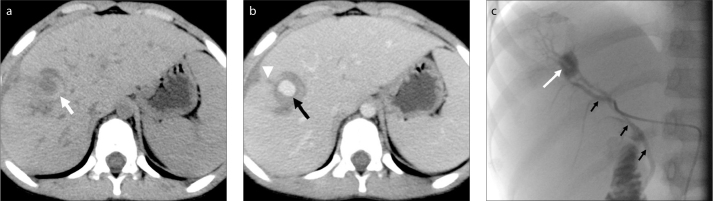
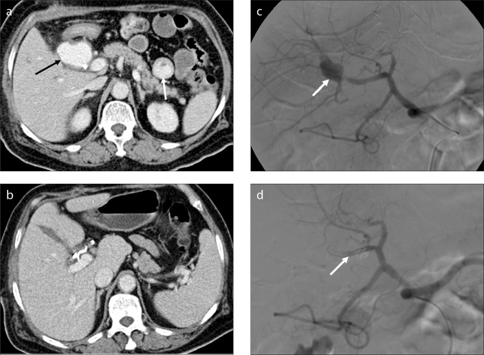
Purpose: We aimed to evaluate the imaging features of computed tomography (CT) and angiography and the efficacy of transcatheter arterial embolization (TAE) in patients with hemobilia of different iatrogenic causes.

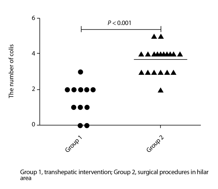
Methods: Thirty patients with hemobilia were divided into two groups according to their iatrogenic causes, i.e., group 1, 11 patients (36.7%) with transhepatic intervention and group 2, 19 patients (63.3%) with surgical procedures in the hilar area. Seventeen patients (56.7%) underwent abdominal contrast-enhanced CT before selective angiography. Polyvinyl alcohol particles, gelatin sponges, and coils were used for TAE. Data from the two groups were compared using Fisher's exact test and the Mann-Whitney U test.

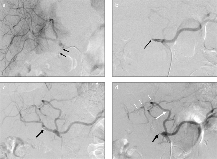
Results: Contrast-enhanced CT showed a hematoma, extravasation of contrast material, and pseudoaneurysm. The bleeding source was determined by angiographic features in all patients, which were not significantly different between the two groups (P = 0.127), and pseudoaneurysm was the most common. The embolic material and number of coils used for TAE were significantly different between the two groups (P < 0.001), but the embolization was technically successful in all patients. The clinical success rate of the first embolization was 100% in group 1 vs. 84.2% in group 2. The overall clinical success rate of TAE was 100% in all patients. The complication rate was 63.6% in group 1 vs. 68.4% in group 2 (P = 1.000).

Conclusion: CT was useful in diagnosing hemobilia, and angiograms enabled determination of the bleeding source. Pseudoaneurysm was one of the most common angiographic features. TAE was successfully performed with different embolic materials on the basis of the iatrogenic cause and bleeding location.


Figures

Figure 1
Figure 1
a–c. A 13-year-old boy with hemobilia after percutaneous transhepatic biliary drainage (PTBD). Unenhanced abdominal CT image (a) shows a hematoma in the hepatic parenchyma (white arrow). Contrast-enhanced abdominal CT image (b) shows enhancement of a pseudoaneurysm (black arrow) near an injury to the hepatic parenchyma due to PTBD (white arrowhead). Selective angiogram of the right hepatic artery (c) shows a pseudoaneurysm in the segment V branch of the right hepatic artery (white arrow) and an arteriobiliary fistula (black arrows).
Figure 2
Figure 2
a–c. A 58-year-old man with hemobilia after PTBD and biliary stent implantation. Unenhanced abdominal CT image (a) shows a hematoma (black arrow) in the gallbladder. Contrast-enhanced abdominal CT image (b) shows a small pseudoaneurysm (black arrow) near a hematoma within the parenchyma (white arrows), and hepatic parenchymal injury due to PTBD (white arrowheads). Superselective angiogram of the right hepatic artery (c) shows a small pseudoaneurysm in the subsegment VIII artery (white arrow) and an arteriobiliary fistula (black arrow).
Figure 3
Figure 3
a–d. A 43-year-old man with hemobilia after laparoscopic cholecystectomy. Contrast-enhanced abdominal CT image (a) shows a large pseudoaneurysm (black arrow) and extravasation of contrast material into the small intestinal cavity (white arrow). Selective angiogram of the right hepatic artery (b) shows a pseudoaneurysm arising from the cystic artery ligation of the right hepatic artery (white arrow). Contrast-enhanced abdominal CT image after TAE (c) shows the disappearance of the pseudoaneurysm and coils in the hilar area. Selective angiogram of the right hepatic artery after embolization with gelatin sponge (1×1×1 mm) and coils (8–50 mm, 5–50 mm) (d) shows the absence of opacification of the pseudoaneurysm (white arrow).
Figure 4
Figure 4
a–d. A 58-year-old man with hemobilia after the resection of cholangiocarcinoma. Selective angiogram of the common hepatic artery (a) shows extravasation of contrast material through the common hepatic artery (black arrows). Selective angiogram after embolization with two coils (8–50 mm) (b) shows the absence of contrast extravasation and occlusion of the common hepatic artery (black arrow). Because of rebleeding three days after the first TAE, the second selective angiogram (c) shows recanalization of the common hepatic artery (black arrow). Selective angiogram of the celiac artery after embolization with coils (5–50 mm, 8–50 mm) (d) shows occlusion of the common hepatic artery (black arrow) and the accessory left hepatic artery (long white arrow) originating from the left gastric artery, which compensates for the blood supply to the hepatic parenchyma through the communicating vessels (short white arrows).
Figure 5
Figure 5
Scatter plot shows the total number of coils used in both groups. The number of coils in group 1 patients with transhepatic intervention is less than that used in group 2 patients with surgical procedures in the hilar area. The difference is significant between the two groups (P < 0.001).

References

    1. Bloechle C, Izbicki JR, Rashed MY, et al. Hemobilia: presentation, diagnosis, and management. Am J Gastroenterol. 1994;89:1537–1540. - PubMed
    1. Murugesan SD, Sathyanesan J, Lakshmanan A, et al. Massive hemobilia: a diagnostic and therapeutic challenge. World J Surgery. 2014;38:1755–1762. http://dx.doi.org/10.1007/s00268-013-2435-5. - DOI - PubMed
    1. Green MHA, Johnson CD, Jamieson NV. Haemobilia. Br J Surg. 2001;88:773–786. http://dx.doi.org/10.1046/j.1365-2168.2001.01756.x. - DOI - PubMed
    1. Yoshida J, Donahue PE, Nyhus LM, et al. Hemobilia: review of recent experience with a worldwide problem. Am J Gastroenterol. 1987;82:448–453. - PubMed
    1. Cao H, Liu J, Li T, et al. Interventional therapy for the treatment of severe hemobilia after percutaneous transhepatic cholangial drainage: a case series. International Surg. 2013;98:223–228. http://dx.doi.org/10.9738/INTSURG-D-13-CC194. - DOI - PMC - PubMed

LinkOut - more resources