Review
. 2016 Jul 1;8(7):a021865.
doi: 10.1101/cshperspect.a021865.

The Discovery and Early Days of TGF-β: A Historical Perspective

Affiliations
Review

The Discovery and Early Days of TGF-β: A Historical Perspective

Harold L Moses et al. Cold Spring Harb Perspect Biol. .

Abstract

Transforming growth factors (TGFs) were discovered as activities that were secreted by cancer cells, and later by normal cells, and had the ability to phenotypically and reversibly transform immortalized fibroblasts. TGF-β distinguished itself from TGF-α because it did not bind to the same epidermal growth factor (EGF) receptor as TGF-α and, therefore, acted through different cell-surface receptors and signaling mediators. This review summarizes the discovery of TGF-β, the early developments in its molecular and biological characterization with its many biological activities in different cell and tissue contexts and its roles in disease, the realization that there is a family of secreted TGF-β-related proteins with many differentiation functions in development and activities in normal cell and tissue physiology, and the subsequent identification and characterization of the receptors and effectors that mediate TGF-β family signaling responses.


Figures

Figure 1.
Figure 1.
Purification to homogeneity of transforming growth factor β (TGF-β) from bovine kidney and human platelets. Human platelets contain ∼100-fold greater amounts of TGF-β than most other normal tissues. The scale of purification from bovine kidneys (or placenta) was large, requiring volumes of solvents unlikely to be used in laboratories today, and requiring four chromatography steps. In contrast, purification of TGF-β to homogeneity from human platelets was achieved using just two chromatography steps on Bio-Gel P-60. These large-scale purification schemes enabled the first determination of the amino-terminal sequence and amino acid composition of TGF-β1. HPCL, High-performance liquid chromatography.
Figure 2.
Figure 2.
Structure and function of pre-pro-transforming growth factor β (TGF-β). (A) Complementary DNA (cDNA) cloning of TGF-β showed that it derives from a large precursor with a signal peptide, the large pro-segment (latency-associated protein [LAP]), and the carboxy-terminal mature biologically active protein (in dimeric form). Large green arrows show processing sites, black bars and asterisks show the position of cysteine residues, and # shows two adjacent cysteine residues. The pattern of cross-linking of the four intrachain disulfides is shown in brackets. Red arrows (down) show the position of the interchain disulfide bridges, whereas a green arrow (up) shows the cysteine that links LAP to latent TGF-β-binding protein (LTBP). A blue underscore shows the position of glycosylation sites and a red underscore shows the position of the Arg-Gly-Asp (RGD)-integrin-binding sequence present in the LAPs of TGF-β1 and TGF-β3 but not the TGF-β2 LAP. (B) TGF-β is secreted from cells in a biologically inactive form (latent TGF-β), with noncovalent association of the dimeric LAP with the dimeric mature carboxy-terminal 112 amino acid TGF-β. Latent TGF-β can be activated by a variety of treatments in vitro resulting in dissociation of the LAP protein from mature TGF-β, thus unmasking its receptor-binding epitopes.
Figure 3.
Figure 3.
Roles of transforming growth factor β (TGF-β) in pathophysiology. TGF-β plays prominent roles in wound healing, fibrosis, and carcinogenesis, as well as a host of other diseases. Listed on the right are various TGF-β-dependent cellular mechanisms that contribute to its effects. ECM, Extracellular matrix.
Figure 4.
Figure 4.
Initial working model of Smad signaling proposed in 1996. Following the identification of Smads as transforming growth factor β (TGF-β) family signaling effectors and several reports on how they might do so, this first model was proposed, based on these results and the perceived analogy with the established mode of signal transducers and activators of transcription (STAT) signaling. (From Derynck and Zhang 1996; reprinted, with permission, from the authors.)
Figure 5.
Figure 5.
Early contributors to the development of the transforming growth factor β (TGF-β) field. The picture taken in February 2006 shows (seated from left) Rik Derynck, Anita Roberts, Harold Moses, and (standing from left) Michael Sporn and Joan Massagué.

References

    1. Akhurst RJ, Derynck R. 2001. TGF-β signaling in cancer—A double-edged sword. Trends Cell Biol 11: S44–S51. - PubMed
    1. Alliston T, Choy L, Ducy P, Karsenty G, Derynck R. 2001. TGF-β-induced repression of CBFA1 by Smad3 decreases cbfa1 and osteocalcin expression and inhibits osteoblast differentiation. EMBO J 20: 2254–2272. - PMC - PubMed
    1. Anzano MA, Roberts AB, Smith JM, Lamb LC, Sporn MB. 1982. Purification by reverse-phase high-performance liquid chromatography of an epidermal growth factor-dependent transforming growth factor. Anal Biochem 125: 217–224. - PubMed
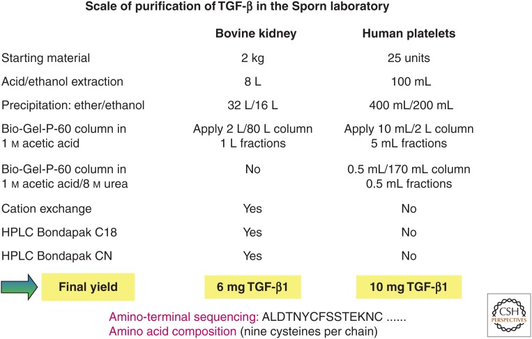
    1. Assoian RK, Komoriya A, Meyers CA, Miller DM, Sporn MB. 1983. Transforming growth factor β in human platelets. Identification of a major storage site, purification, and characterization. J Biol Chem 258: 7155–7160. - PubMed
    1. Attisano L, Wrana JL, Cheifetz S, Massagué J. 1992. Novel activin receptors: Distinct genes and alternative mRNA splicing generate a repertoire of serine/threonine kinase receptors. Cell 68: 97–108. - PubMed

MeSH terms

Substances