. 2016 Jun 22:6:28512.
doi: 10.1038/srep28512.

R-spondin 2 promotes acetylcholine receptor clustering at the neuromuscular junction via Lgr5

Affiliations

R-spondin 2 promotes acetylcholine receptor clustering at the neuromuscular junction via Lgr5

Hiroaki Nakashima et al. Sci Rep. .

Abstract

At the neuromuscular junction (NMJ), acetylcholine receptor (AChR) clustering is mediated by spinal motor neuron (SMN)-derived agrin and its receptors on the muscle, the low-density lipoprotein receptor-related protein 4 (LRP4) and muscle-specific receptor tyrosine kinase (MuSK). Additionally, AChR clustering is mediated by the components of the Wnt pathway. Laser capture microdissection of SMNs revealed that a secreted activator of Wnt signaling, R-spondin 2 (Rspo2), is highly expressed in SMNs. We found that Rspo2 is enriched at the NMJ, and that Rspo2 induces MuSK phosphorylation and AChR clustering. Rspo2 requires Wnt ligands, but not agrin, for promoting AChR clustering in cultured myotubes. Leucine-rich repeat-containing G-protein coupled receptor 5 (Lgr5), an Rspo2 receptor, is also accumulated at the NMJ, and is associated with MuSK via LRP4. Lgr5 is required for Rspo2-mediated AChR clustering in myotubes. In Rspo2-knockout mice, the number and density of AChRs at the NMJ are reduced. The Rspo2-knockout diaphragm has an altered ultrastructure with widened synaptic clefts and sparse synaptic vesicles. Frequency of miniature endplate currents is markedly reduced in Rspo2-knockout mice. To conclude, we demonstrate that Rspo2 and its receptor Lgr5 are Wnt-dependent and agrin-independent regulators of AChR clustering at the NMJ.


Figures

Figure 1
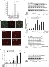
Figure 1. R-spondin 2 (Rspo2) is highly expressed in laser capture microdissection-harvested spinal motor neurons (SMNs) of the mouse spinal cord.
(A) Toluidine blue-stained section of the cervical spinal cord of a 6-week-old C57BL6/J mouse before laser capture microdissection. Arrows indicate SMNs to be dissected. (B) The left anterior horn region (enlarged from A) after the dissection of SMNs. Orange lines mark the traces of the laser beam. (C) The right posterior horn region (enlarged from A) after the dissection of posterior horn cells. Orange line marks the trace of the laser beam. (D) A representative dissected SMN. (E) The ratio of mRNA expressions in SMNs and posterior horn cells of Agrn, Chat, Isl1, Mnx1, and Wnt-related genes (Rspo, Wnt, Fzd, and Lrp genes) according to the Affymetrix microarray data. Agrn, Chat, Isl1, Mnx1, Fzd, and Lrp encode agrin, choline acetyltransferase, islet-1, HB9, frizzled, and low-density lipoprotein receptor-related protein, respectively. (F) In situ hybridization of Rspo2 in the cervical spinal cord of a 6-week-old C57BL6/J mouse.
Figure 2
Figure 2. Rspo2 is enriched at the NMJ and activates MuSK to induce AChR clustering.
(A) Rspo2 expression in the diaphragm and spinal cord normalized by Gapdh and also to E18.5 diaphragm. Mean and SD (n = 3) are indicated. (B) Rspo2 immunostaining and α-bungarotoxin staining for AChR at the NMJ of the tibialis anterior muscle (cross section). (C) AChR clusters visualized with α-bungarotoxin (red). C2C12 myotubes were cultured with 0.05 nM agrin and/or 0.05 nM Rspo2. Arrows point to the AChR clusters with an axis length of 4 μm or more. Blinded morphometric analysis is shown in Supplementary Fig. S2A. (D) ATF2-luciferase reporter assay to quantify agrin (10 ng/ml)- and Rspo2 (100 ng/ml)-mediated activation of MuSK signaling in transfected HEK293 cells. Relative luciferase activities (RLA) are normalized to that with empty vectors. Mean and SD are indicated (n = 3). **p < 0.01 by t-test. n.s., no significant difference. (E) Agrin- and Rspo2-mediated MuSK phosphorylation in C2C12 myotubes. Phosphorylated MuSK was immunoprecipitated (IP) and immunoblotted (IB) with indicated antibodies. (F) The ratio of phosphorylated (phosphoMuSK) and total MuSK was normalized to that with 0.1 nM agrin. Mean and SD (n = 3) are indicated. **p < 0.01 by t-test. The mean is also indicated in (E). BSA was added to control the amount of total proteins. (G) Additive effect of 0.05 nM agrin and 0.05 nM Rspo2 on MuSK phosphorylation in C2C12 myotubes. Phosphorylated MuSK was detected as in (E). Band intensities were normalized to that with 0.1 nM agrin (Supplementary Fig. S2B). The mean intensity is also is indicated below the blot. BSA was added as in (E). (H) Agrin- and Rspo2-mediated expression of rapsyn in C2C12 myotubes. Band intensities were normalized as in (G) (Supplementary Fig. S2C). The mean value is also indicated below the blot.
Figure 3
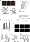
Figure 3. The Rspo2/Lgr5 complex induces activation and phosphorylation of MuSK and AChR clustering.
(A) Lgr5 immunostaining and α-bungarotoxin staining of the NMJs as in Fig. 2B. (B,C) Lgr5 was co-immunoprecipitated with anti-myc (B) or anti-Flag (C) antibody in HEK293 cells. The normalized ratio of co-immunoprecipitated Lgr5 to total Lgr5 is shown in Supplementary Fig. S3A and S3B, and the mean value is indicated below the blot. (D) ATF2-luciferase reporter assays as in Fig. 2D in siRNA-transfected HEK293 cells. RLA are normalized to that of BSA with siControl. Mean and SD are indicated (**p < 0.01 by t-test, n = 3). Efficiency of siLgr5 and the rescue experiment is shown in Supplementary Figs S3B and S3C, respectively. (E) The effects of Lgr5 on MuSK phosphorylation in L cells treated with 0.1 nM Rspo2. Total Flag-MuSK was immunoprecipitated (IP), and immunoblotted (IB) with indicated antibodies. The ratio of phosphorylated and total Flag-MuSK was normalized to that of the control knockdown (shControl) in lane 3. Quantification is shown in Supplementary Fig. S3F, and the mean value is indicated below the blot. Efficiency of shLgr5 in L cells is indicated in Supplementary Fig. S3E. (F) C2C12 myoblasts were infected with lentivirus expressing shControl or shLgr5. After differentiation into myotubes, 0.1 nM of BSA (Control), agrin, or Rspo2 was added. Phosphorylated MuSK was immunoprecipitated (IP) and immunoblotted (IB) with indicated antibodies. The band intensities were normalized to that of cells treated with 0.1 nM agrin and shControl. Quantification of phosphorylated MuSK is shown in Supplementary Fig. S3H, and the mean value is indicted below the blot. Efficiency of shLgr5 in C2C12 myotubes is indicated in Supplementary Fig. S3G. (G,H) C2C12 myoblasts were infected with lentivirus expressing shControl or shLgr5. After differentiation into myotubes, 0.1 nM of BSA (Control), agrin, or Rspo2 was added. AChR cluster was visualized with α-bungarotoxin (red). (F) Arrows point to AChR clusters in representative images. (G) Blinded morphometric analysis. Mean and SD are indicated (**p < 0.01 by t-test, n = 3). Instead of purified recombinant Rspo2, we also used Rspo2-containing conditioned medium, and showed the results in Supplementary Fig. S2D.
Figure 4
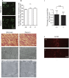
Figure 4. Lack of R-spondin 2 (Rspo2) in mice has minimal effects on spinal motor neuron (SMN) survival and muscle differentiation, but has a significant effect on acetylcholine receptor (AChR) clusters in the left diaphragm.
(A) Immunostaining for islet1/2 expressed in the SMNs of the spinal cord (C3-C6) at embryonic day (E) 18.5. (B) The number of islet1/2-positive SMNs in wild-type (+/+), heterozygous Rspo2-knockout (+/−), and homozygous Rspo2-knockout (−/−) mice. Bars indicate the mean and standard error of mean (SE) (n > 90). No statistically significant differences (n.s.) were observed with one-way ANOVA. (C,D) Hematoxylin and eosin staining of the tibialis anterior muscle of mice at E18.5. (E–H) Representative electron micrographs of the diaphragms at E18.5 at different magnifications. (I) Thickness of muscle fibers at the Z disk in the left diaphragms of wild-type (+/+) and Rspo2-knockout (−/−) mice. Five to seven electron micrographs were analyzed in each mouse. (J) Surface views of the left diaphragms harvested from wild-type (+/+) and Rspo2-knockout (−/−) mice at E18.5. AChR was stained with Alexa546-conjugated α-bungarotoxin (red). The widths of the AChR bands of wild-type and Rspo2-kockout diaphragms were 143.34 ± 3.73 μm and 221.85 ± 6.52 μm, respectively (p < 0.0001 by Student’s t-test, n = 5).
Figure 5
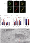
Figure 5. Apposition of nerve terminals and muscle endplates is compromised in the E18.5 diaphragm of R-spondin 2 (Rspo2)−/− mice.
(A–F) Representative confocal images of the left diaphragm at E18.5 labeled with an anti-synaptophysin antibody and α-bungarotoxin to visualize the nerve terminals and acetylcholine receptors (AChRs), respectively. Endplates of the wild-type muscles were mostly ovoid-shaped (B), whereas the endplates of Rspo2−/− muscles were large, round, and heterogeneously stained (E). (G–I) Blinded morphometric analysis of synaptophysin (G) and AChR (H) in the AChR clusters revealed that NMJ areas were markedly enlarged at E18.5. Numbers of synaptophysin-positive clusters (G) and AChR-positive clusters (H) are shown. (I) The ratio is calculated by dividing the synaptophysin-positive area by the AChR-positive area. Note that not all AChR-positive (red) pixels were synaptophysin-positive (green) in each AChR cluster. Mean and standard deviation (SD; n = 6) are indicated. ****p < 0.001, ***p < 0.005, and *p < 0.05 by t-test. n.s., no significant difference. (J,K) Representative electron micrographs of the neuromuscular junctions (NMJs) in the diaphragm of wild-type and Rspo2−/− mice at E18.5. The red two-headed arrow indicates a widened synaptic cleft. The closed arrowhead at wild-type endplate points to a postsynaptic fold. In the Rspo2−/− mice, synaptic vesicles were larger and sparser than those in wild-type mice. SV, synaptic vesicles; TS, terminal Schwann cell. Low magnification images are shown in Supplementary Fig. S6A. Blinded morphometric measurements are shown in Table 1.
Figure 6
Figure 6. Schematic image showing the R-spondin 2 (Rspo2)-mediated acetylcholine receptor (AChR) clustering at the neuromuscular junction (NMJ).
Rspo2 binds to leucine-rich repeat-containing G-protein coupled receptor 5 (Lgr5) on the endplate and phosphorylates muscle-specific receptor tyrosine kinase (MuSK) to induce AChR clustering in an agrin-independent manner.

Similar articles

Cited by

References

    1. Burden S. J. SnapShot: Neuromuscular Junction. Cell 144, 826–826 e821 (2011). - PubMed
    1. Wu H., Xiong W. C. & Mei L. To build a synapse: signaling pathways in neuromuscular junction assembly. Development 137, 1017–1033 (2010). - PMC - PubMed
    1. Sanes J. R. & Lichtman J. W. Induction, assembly, maturation and maintenance of a postsynaptic apparatus. Nat Rev Neurosci 2, 791–805 (2001). - PubMed
    1. Bezakova G. & Ruegg M. A. New insights into the roles of agrin. Nat Rev Mol Cell Biol 4, 295–308 (2003). - PubMed
    1. McMahan U. J. et al.. Agrin isoforms and their role in synaptogenesis. Curr Opin Cell Biol 4, 869–874 (1992). - PubMed

Publication types