

Longitudinal diffusion tensor imaging after pediatric traumatic brain injury: Impact of age at injury and time since injury on pathway integrity
- PMID: 27329317
- PMCID: PMC5053864
- DOI: 10.1002/hbm.23286
Longitudinal diffusion tensor imaging after pediatric traumatic brain injury: Impact of age at injury and time since injury on pathway integrity
Abstract
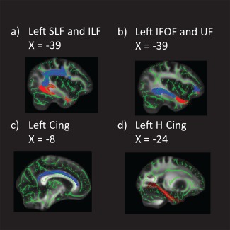
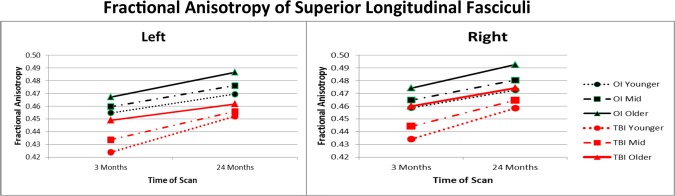
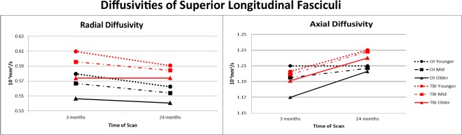
Following pediatric traumatic brain injury (TBI), longitudinal diffusion tensor imaging may characterize alterations in initial recovery and subsequent trajectory of white matter development. Our primary aim examined effects of age at injury and time since injury on pathway microstructure in children ages 6-15 scanned 3 and 24 months after TBI. Microstructural values generated using tract-based spatial statistics extracted from core association, limbic, and projection pathways were analyzed using general linear mixed models. Relative to children with orthopedic injury, the TBI group had lower fractional anisotropy (FA) bilaterally in all seven pathways. In left-hemisphere association pathways, school-aged children with TBI had the lowest initial pathway integrity and showed the greatest increase in FA over time suggesting continued development despite incomplete recovery. Adolescents showed limited change in FA and radial diffusivity and had the greatest residual deficit suggesting relatively arrested development. Radial diffusivity was persistently elevated in the TBI group, implicating dysmyelination as a core contributor to chronic post-traumatic neurodegenerative changes. The secondary aim compared FA values over time in the total sample, including participants contributing either one or two scans to the analysis, to the longitudinal cases contributing two scans. For each pathway, FA values and effect sizes were very similar and indicated extremely small differences in measurement of change over time in the total and longitudinal samples. Statistical approaches incorporating missing data may reliably estimate the effects of TBI and provide increased power to identify whether pathways show neurodegeneration, arrested development, or continued growth following pediatric TBI. Hum Brain Mapp 37:3929-3945, 2016. © 2016 Wiley Periodicals, Inc.
Keywords: axial diffusivity; children; chronic; dysmyelination; fractional anisotropy; microstructure; neurodegeneration; radial diffusivity; tract-based spatial statistics.
© 2016 Wiley Periodicals, Inc.
Figures
References
-
- Anderson V, Catroppa C, Morse S, Haritou F, Rosenfeld J (2005): Functional plasticity or vulnerability after early brain injury? Pediatrics 116:1374–1382. - PubMed
-
- Andersson JLR, Jenkinson M, Smith S (2007): Non‐linear registration aka spatial normalization. Oxford UK: FMRIB Centre. Report nr TR07JA2.
Publication types
MeSH terms
Grants and funding
LinkOut - more resources
Full Text Sources
Other Literature Sources
Medical
