. 2016 Jun 22:7:11929.
doi: 10.1038/ncomms11929.

ASC filament formation serves as a signal amplification mechanism for inflammasomes

Affiliations

ASC filament formation serves as a signal amplification mechanism for inflammasomes

Mathias S Dick et al. Nat Commun. .

Erratum in

Abstract

A hallmark of inflammasome activation is the ASC speck, a micrometre-sized structure formed by the inflammasome adaptor protein ASC (apoptosis-associated speck-like protein containing a CARD), which consists of a pyrin domain (PYD) and a caspase recruitment domain (CARD). Here we show that assembly of the ASC speck involves oligomerization of ASC(PYD) into filaments and cross-linking of these filaments by ASC(CARD). ASC mutants with a non-functional CARD only assemble filaments but not specks, and moreover disrupt endogenous specks in primary macrophages. Systematic site-directed mutagenesis of ASC(PYD) is used to identify oligomerization-deficient ASC mutants and demonstrate that ASC speck formation is required for efficient processing of IL-1β, but dispensable for gasdermin-D cleavage and pyroptosis induction. Our results suggest that the oligomerization of ASC creates a multitude of potential caspase-1 activation sites, thus serving as a signal amplification mechanism for inflammasome-mediated cytokine production.


Figures
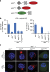
Figure 1
Figure 1. Both domains of ASC are required for signalling.
(a) Schematic representation of the domain organization of fluorophore-tagged (mCherry or enhanced GFP (eGFP)) WT ASC (ASCFL), ASCPYD and ASCCARD constructs. (b) Release of LDH (assessing cell death) and IL-1β from LPS-primed immortalized WT, Asc−/− or Asc−/− BMDMs expressing ASCFL, ASCPYD or ASCCARD 3 h after poly(dA:dT) transfection (1 μg ml−1). (c) Representative images of cell lines from (b) 3 h after poly(dA:dT) transfection (1 μg ml−1) or 1 h after ATP treatment (5 mM). DNA (blue, Hoechst), ASCFL or ASCPYD (red) and ASCCARD (green). Scale bars, 10 μm. Data (b,c) are representative of three independent experiments. Graphs show the mean and s.d. from quadruplicate wells. See also Supplementary Fig. 1.
Figure 2
Figure 2. The CARD of ASC condenses PYD filaments into the speck.
(a) Structural model of the mouse ASCCARD based on the human homologue (PDB 2KN6 (ref. 67)). The structure is shown in ribbon (left) and electrostatic surface representation (right, blue, positive charge; red, negative charge). Residues D130 and D134, involved in the interaction with pro-caspase-1, are highlighted. (b) Release of LDH and IL-1β from LPS-primed immortalized Asc−/− BMDMs and Asc−/−BMDMs expressing ASCFL, ASCD130R or ASCD134R 3 h after poly(dA:dT) transfection (1 μg ml−1). (c,d) Representative images of cell lines from (b) 3 h after poly(dA:dT) transfection (1 μg ml−1) (c) or 1 h after ATP treatment (5 mM) (d). DNA was stained with Hoechst (blue) and ASC (red). Scale bars, 10 μm. (e,f) Measurement of the ASC speck diameter in primary C57BL/6 BMDMs transduced with the indicated ratio of mCherry-tagged ASCFL or ASCD130R and transfected with poly(dA:dT) (3 h at 1 μg ml−1 (e)) or treated with nigericin (1 h, 20 μM (f)) after LPS priming. (g) Representative images from e. DNA was stained with Hoechst (blue) and ASC (red). Scale bars, 5 μm. Data are representative of three (bd) independent experiments. Graphs show the mean and s.d. from quadruplicate wells (b) or triplicate coverslips (e,f). The numbers of specks measured were 99, 92, 104, 94 and 108 in (e) and 149, 134, 85, 98 and 95 in (f). *P<0.05, **P<0.01 and ***P<0.001 (one-way analysis of variance). See also Supplementary Figs 2 and 3.
Figure 3
Figure 3. A bridging ASC molecule is required for ASC speck formation after NLRC4 activation.
(a) Release of LDH and IL-1β from LPS-primed immortalized WT, Asc−/− or Asc−/− BMDMs expressing ASCFL, ASCPYD or ASCCARD after infection with log-phase WT S. Typhimurium SL1344 (multiplicity of infection (MOI) 10 for 1 h). (b) Quantification of the percentage of cells from a with ASC specks or filaments (collectively referred to as ASC aggregates). (c) Representative images of cell lines from b. DNA (blue, Hoechst) and ASC (red). Scale bars, 10 μm. (d) Release of LDH and IL-1β from LPS-primed Asc−/− BMDMs and Asc−/− BMDMs expressing ASCFL, ASCD130R or ASCD134R after infection with log-phase WT S. Typhimurium SL1344 (MOI 10 for 1 h). (e) Quantification of the percentage of cells with ASC aggregates from d. (f) Measurement of the ASC speck diameter in primary C57BL/6 BMDMs transduced with the indicated ratio of mCherry-tagged ASCFL or ASCD130R and infected with log-phase WT S. Typhimurium SL1344 (MOI 10 for 1 h) after LPS priming. Data are representative of three (ae) independent experiments. Graphs show the mean and s.d. from quadruplicate wells in (a,ce) and triplicate coverslips in f. The numbers of specks measured were 194, 134, 184, 128 and 141 in f. *P<0.05, **P<0.01 and ***P<0.001 (one-way analysis of variance). See also Supplementary Fig. 3.
Figure 4
Figure 4. Mutations in interface II uncouple speck formation and IL-1β release from cell death.
(a) Detailed view of the three interaction interfaces forming the ASCPYD filament (PDB 2N1F (ref. 15), shown in Supplementary Fig. 4a). The polypeptide backbones are shown in ribbon representation. All amino acid side chains involved in intersubunit contacts are shown as stick models. Residues mutated in this study are coloured red with their sequence label. (bd) Quantification of ASC aggregates or the release of LDH and IL-1β from LPS-primed immortalized Asc−/− BMDMs and Asc−/− BMDMs expressing ASCFL or the indicated ASC mutants 3 h after poly(dA:dT) transfection (1 μg ml−1). ASCY59A and ASCE80R are highlighted in grey. Graphs show means and s.d. from quadruplicate wells or ten random fields of view. Data are representative of at least three independent experiments. See also Supplementary Figs 4 and 6.
Figure 5
Figure 5. ASC–receptor interaction and ASC filament formation can be uncoupled genetically.
(a) Western blot analysis of the interaction of AIM2 with ASCFL or the indicated ASC mutants. AIM2-V5 was immunoprecipitated from lysates of HEK293T cells co-transfected with AIM2-V5 and the indicated ASC mutants. Co-immunoprecipitating proteins were identified using anti-ASC and anti-V5. *Immunoglobulin heavy chain. Results shown are representative from two independent experiments. (b) Filament formation of WT ASCPYD and its single amino-acid variants K21A, Y59A and E80R in vitro monitored by dynamic light scattering. Normalized growth signals (IN) are reported as a function of time for one representative experiment for each variant (dots). Best fits to single exponential functions are shown with solid lines. (c) Kinetic rate constants kF of filament formation obtained from three independent experiments. (d) Representative negative-stained TEM micrographs of filament formed by ASCPYD and its variants after 350 min of incubation at physiological pH condition. Scale bars, 200 nm. See also Supplementary Figs 7 and 9.
Figure 6
Figure 6. Caspase-1 but not gasdermin-D processing depends on ASC oligomerization.
(a) Western blot analysis for cleaved caspase-1 p20, IL-1β p17, and HMGB-1 in cell supernatants (SN) and pro-caspase-1, pro-IL-1β and HMGB-1 in cell lysates (lys) of LPS-primed immortalized Asc−/− BMDMs or Asc−/− BMDMs expressing ASCFL, ASCY59A or ASCE80R 3 h after poly(dA:dT) transfection (1 μg ml−1). (b) Release of LDH from LPS-primed immortalized Asc−/− BMDMs or Asc−/− BMDMs expressing ASCFL, ASCY59A or ASCE80R, or derived Casp1 knockouts 3 h after poly(dA:dT) transfection (1 μg ml−1). (c) Western blot analysis for processing of full-length gasdermin-D (GSDMDFL) into the active N-terminal fragment (GSDMDN-term) in combined lysates and supernatants (lys + SN) of LPS-primed immortalized Asc−/− BMDMs expressing ASCFL, ASCY59A or ASCE80R, or derived Casp1 knockouts 3 h after poly(dA:dT) transfection (1 μg ml−1). Arrowhead, gasdermin-DNterm p30; *a cross-reacting band. (d) Release of LDH from LPS-primed primary C57BL/6 WT (WT), Casp1−/−/Casp11−/− or Asc−/− BMDMs infected with S. Typhimurium (multiplicity of infection (MOI)=10, 1 h). (e) Western blot analysis for processing of full-length gasdermin-D (GSDMDFL) into the active N-terminal fragment (GSDMDN-term) in combined lysates and supernatants (lys+SN) of LPS-primed primary C57BL/6 WT (WT), Casp1−/−/Casp11−/− or Asc−/− BMDMs infected with S. Typhimurium (MOI=10, 1 h) or left uninfected. Arrowhead, gasdermin-DNterm p30; *a cross-reacting band; ** a S. Typhimurium-specific cross-reactive band. See also Supplementary Figs 8 and 9.
Figure 7
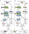
Figure 7. Model of signal amplification by ASC filaments.
(a) CARD-containing receptors recruit the adaptor protein ASC via homotypic CARD–CARD interactions, that is, a bridging ASC molecule. This step nucleates the ASCPYD of several bridging ASC molecules, leading to the formation of an ASCPYD filament, which is condensed into the ASC speck by the ASCCARD. Filament formation promotes the activation of large quantities of caspase-1, thus promoting the proteolytic maturation of large amounts of cytokines (pro-IL-1β). In the absence of ASC, CARD-containing receptors directly interact with pro-caspase-1, leading to the formation of so-called ‘death complexes'. In these small complexes, only few molecules of caspase-1 are activated and pro-caspase-1 processing might not happen. The few molecules of caspase-1 are sufficient to effectively induce pyroptosis, but cytokine processing is reduced. (b) PYD-containing receptors can directly interact with ASC via homotypic PYD–PYD interactions, leading to ASCPYD filaments and finally the ASC speck. As for CARD-containing receptors, this leads to caspase-1 activation and subsequent cytokine processing and pyroptosis. Mutations blocking or slowing ASC filament formation (for example, ASCE80R or ASCY59A) only allow for few molecules of caspase-1 being activated. This is sufficient to induce pyroptosis, but insufficient to produce large amounts of mature cytokines before the cell lyses.

References

    1. von Moltke J., Ayres J. S., Kofoed E. M., Chavarría-Smith J. & Vance R. E. Recognition of bacteria by inflammasomes. Annu. Rev. Immunol. 31, 73–106 (2013). - PubMed
    1. Philpott D. J., Sorbara M. T., Robertson S. J., Croitoru K. & Girardin S. E. NOD proteins: regulators of inflammation in health and disease. Nat. Rev. Immunol. 14, 9–23 (2014). - PubMed
    1. Franchi L., Warner N., Viani K. & Nuñez G. Function of Nod-like receptors in microbial recognition and host defense. Immunol. Rev. 227, 106–128 (2009). - PMC - PubMed
    1. Martinon F., Burns K. & Tschopp J. The inflammasome: a molecular platform triggering activation of inflammatory caspases and processing of proIL-beta. Mol. Cell 10, 417–426 (2002). - PubMed
    1. Schroder K. & Tschopp J. The inflammasomes. Cell 140, 821–832 (2010). - PubMed

Publication types

MeSH terms

Substances