. 2016 Jun 22:7:12070.
doi: 10.1038/ncomms12070.

Structural basis of suppression of host translation termination by Moloney Murine Leukemia Virus

Affiliations

Structural basis of suppression of host translation termination by Moloney Murine Leukemia Virus

Xuhua Tang et al. Nat Commun. .

Abstract

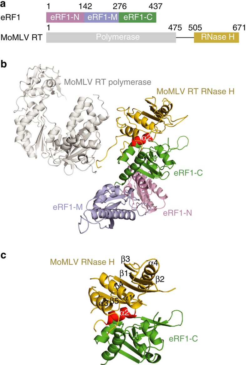
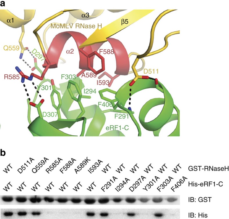
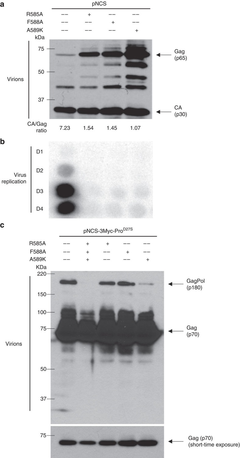
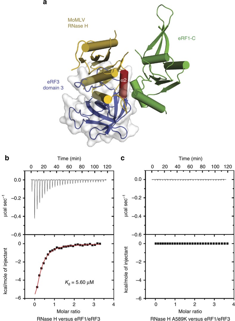
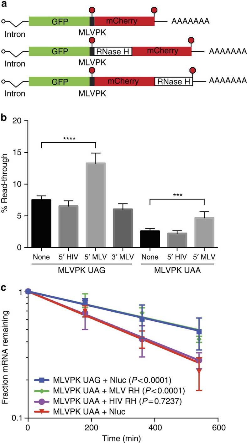
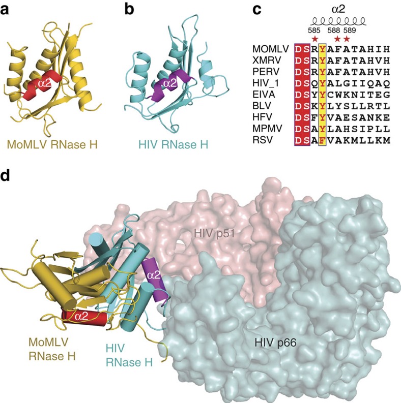
Retroviral reverse transcriptase (RT) of Moloney murine leukemia virus (MoMLV) is expressed in the form of a large Gag-Pol precursor protein by suppression of translational termination in which the maximal efficiency of stop codon read-through depends on the interaction between MoMLV RT and peptidyl release factor 1 (eRF1). Here, we report the crystal structure of MoMLV RT in complex with eRF1. The MoMLV RT interacts with the C-terminal domain of eRF1 via its RNase H domain to sterically occlude the binding of peptidyl release factor 3 (eRF3) to eRF1. Promotion of read-through by MoMLV RNase H prevents nonsense-mediated mRNA decay (NMD) of mRNAs. Comparison of our structure with that of HIV RT explains why HIV RT cannot interact with eRF1. Our results provide a mechanistic view of how MoMLV manipulates the host translation termination machinery for the synthesis of its own proteins.


Figures

Figure 1
Figure 1. Structure of the MoMLV RT/eRF1 complex.
(a) Schematic representation of the domain organization of eRF1 and MoMLV RT. Domains N, M, and C of eRF1 are coloured in pink, lightblue and green, respectively. MoMLV RT polymerase domain is coloured in grey and RNase H domain in yellow. (b) A ribbon diagram of the MoMLV RT/eRF1 complex. The colouring scheme is as in a. The helix α2 of RNase H domain is highlighted in red. (c) The RNase H/eRF1-C complex structure. The secondary structure elements of RNase H domain are labelled.
Figure 2
Figure 2. Interaction of MoMLV RT with eRF1.
(a) Interface between the RNase H domain of MoMLV RT and eRF1-C. Residues involved in the interaction are shown as sticks and labelled. (b) GST pull-down assay. GST tagged WT MoMLV RNase H and its mutants on beads were used to bind His-tagged eRF1-C and its variants.
Figure 3
Figure 3. Non-interacting RT mutants showing reduced read-through are replication defective.
(a) The provirus carrying mutants that are unable to bind eRF1 show reduced read-through efficiency. The proviral DNA were used to transform 293 T cells and the purified virions were analyzed by western blot. The virion produced by WT DNA contained high levels of capsid (CA, ∼30 kDa), while mutants showed poor Gag (∼65 KDa) cleavage as a consequence of low read-through. (b) Non-interacting RT mutants are replication defective. Release of spreading virus was detected on the indicated day post infection. (c) HEK 293 T cells were transfected with WT pNCS-3Myc-ProD27S or pNCS-3Myc-ProD27S bearing indicated mutations. Forty-eight hour after transfection, the viruses in the supernatant were collected and purified by ultra-centrifuge through the 25% sucrose cushion. The virus pellets were re-suspended in protein loading buffer, resolved in SDS–PAGE, and visualized by western blot using c-Myc antibody (9E10; sc-40, Santa Cruz; 1:1000 dilution). Noted that the Gag protein has a molecular mass of ∼70 kDa as carrying the 3Myc tag.
Figure 4
Figure 4. The RNase H domain of MoMLV RT outcompetes eRF3 for binding to eRF1.
(a) Superposition of the RNase H/eRF1-C complex with the eRF1/eRF3 complex (PDB accession code: 3E1Y) at eRF1-C domain. The overlapping interface suggests that MoMLV RT and eRF3 are mutually exclusive for binding to eRF1. eRF3 domain 3 is shown in cartoon (blue) covered with grey transparent surface. eRF3 domain 2 and eRF1-C domain in eRF1/eRF3 complex are not displayed for clarity. (b,c) Representative ITC titrations of WT RNase H and mutant A589K to the eRF1/eRF3 complex. The upper panels show the binding isotherms and the lower panels show the integrated heat for each injection fitted to a single-site model.
Figure 5
Figure 5. MoMLV RNase H enhances stop codon read-through and stabilizes mRNAs.
(a) Schematic of tet-regulated reporter mRNAs used in read-through and mRNA decay assays. The indicated RNase H variants were inserted downstream of the MLVPK, in-frame with the GFP and mCherry ORFs. The red dot represents the stop codon. (b) Translational read-through assays using dual-fluorescent-protein reporters containing the indicated MLVPK and RNase H variants. The ratio of mCherry:GFP from each experimental construct was normalized to that arising from a sequence-matched control lacking a stop codon between GFP and mCherry. Error bars indicate s.d. (n=7; ****P<0.0001; ***P<0.001 in one-way ANOVA with Sidak's multiple comparisons test). (c) Semi-log plot of levels of the indicated tet-regulated mRNAs throughout pulse-chase mRNA decay assays. Best-fit lines calculated by the least-squares method are indicated; error bars denote s.d. (n=4; P values were calculated using ANCOVA analysis, comparing the indicated mRNAs to MLVPK UAA+Nluc).
Figure 6
Figure 6. Structural explanation of why HIV RT cannot interact with eRF1.
(a) Cartoon representation of the RNase H domain of MoMLV RT. (b) The RNase H domain of HIV RT, which is coloured in cyan with its helix α2 highlighted in purple. (c) Sequence alignment of the helix α2 from various genera of retroviruses. MoMLV, XMRV and PERV (Porcine endogenous retrovirus) belong to gammaretrovirus. HIV-1 and EIVA (Equine infectious anaemia) are lentivirus. BLV (Bovine leukemia virus), HFV (Human foamy virus), MPMV (Mason pfizer monkey virus) and RSV (Rous sarcoma virus) belong to deltaretrovirus, spumaretrovirus, betaretrovirus and alpharetrovirus respectively. (d) Superposition of MoMLV RT and HIV RT p66 (PDB accession code: 1HYS) at their polymerase domains. HIV RT p66 except RNase H domain and p51 subunits are shown as surfaces coloured in cyan and salmon, respectively.

Similar articles

Cited by

References

    1. Walsh D. & Mohr I. Viral subversion of the host protein synthesis machinery. Nat. Rev. Microbiol. 9, 860–875 (2011). - PMC - PubMed
    1. Shehu-Xhilaga M., Crowe S. M. & Mak J. Maintenance of the Gag/Gag–Pol ratio is important for human immunodeficiency virus type 1 RNA dimerization and viral infectivity. J. Virol. 75, 1834–1841 (2001). - PMC - PubMed
    1. Felsenstein K. M. & Goff S. P. Expression of the gag-pol fusion protein of Moloney murine leukemia virus without gag protein does not induce virion formation or proteolytic processing. J. Virol. 62, 2179–2182 (1988). - PMC - PubMed
    1. Feng Y. X., Yuan H., Rein A. & Levin J. G. Bipartite signal for read-through suppression in murine leukemia virus mRNA: an eight-nucleotide purine-rich sequence immediately downstream of the gag termination codon followed by an RNA pseudoknot. J. Virol. 66, 5127–5132 (1992). - PMC - PubMed
    1. Felsenstein K. M. & Goff S. P. Mutational analysis of the gag-pol junction of Moloney murine leukemia virus: requirements for expression of the gag-pol fusion protein. J. Virol. 66, 6601–6608 (1992). - PMC - PubMed

Publication types

MeSH terms