. 2016 Jun 22:6:28361.
doi: 10.1038/srep28361.

NdhV subunit regulates the activity of type-1 NAD(P)H dehydrogenase under high light conditions in cyanobacterium Synechocystis sp. PCC 6803

Affiliations

NdhV subunit regulates the activity of type-1 NAD(P)H dehydrogenase under high light conditions in cyanobacterium Synechocystis sp. PCC 6803

Xin Chen et al. Sci Rep. .

Abstract

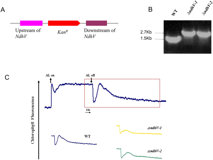
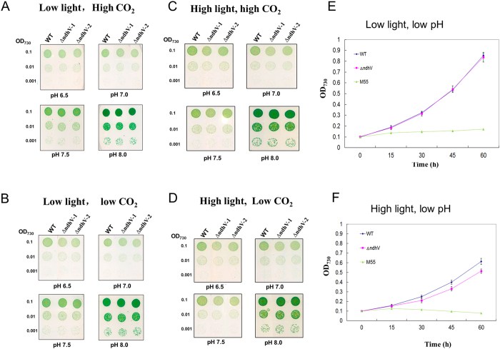
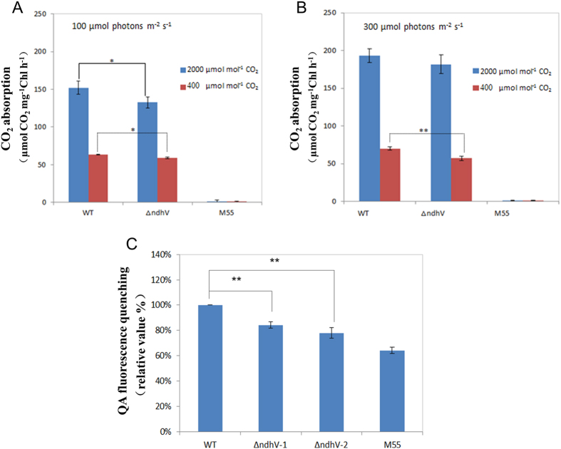
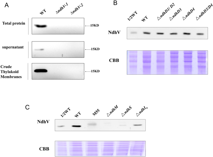
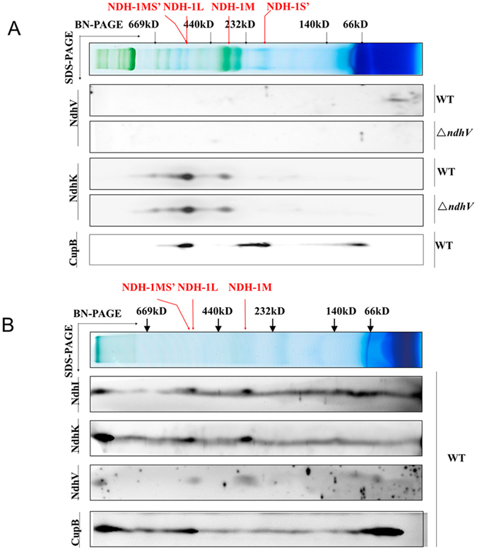
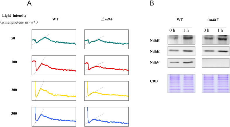
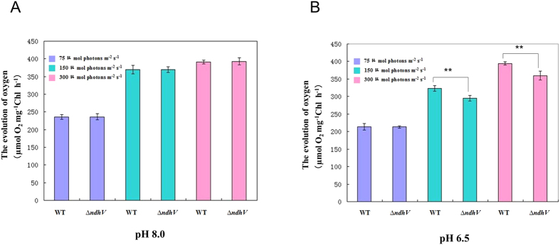
The cyanobacterial NAD(P)H dehydrogenase (NDH-1) complexes play crucial roles in variety of bioenergetic reactions. However, the regulative mechanism of NDH-1 under stressed conditions is still unclear. In this study, we detected that the NDH-1 activity is partially impaired, but the accumulation of NDH-1 complexes was little affected in the NdhV deleted mutant (ΔndhV) at low light in cyanobacterium Synechocystis sp. PCC 6803. ΔndhV grew normally at low light but slowly at high light under inorganic carbon limitation conditions (low pH or low CO2), meanwhile the activity of CO2 uptake was evidently lowered than wild type even at pH 8.0. The accumulation of NdhV in thylakoids strictly relies on the presence of the hydrophilic subcomplex of NDH-1. Furthermore, NdhV was co-located with hydrophilic subunits of NDH-1 loosely associated with the NDH-1L, NDH-1MS' and NDH-1M complexes. The level of the NdhV was significantly increased at high light and deletion of NdhV suppressed the up-regulation of NDH-1 activity, causing the lowered the photosynthetic oxygen evolution at pH 6.5 and high light. These data indicate that NdhV is an intrinsic subunit of hydrophilic subcomplex of NDH-1, required for efficient operation of cyclic electron transport around photosystem I and CO2 uptake at high lights.


Conflict of interest statement

The authors declare no competing financial interests.

Figures

Figure 1
Figure 1. NdhV gene deletion and its effect on NDH-1 activity.
(A) Construction of plasmid to generate NdhV deleted mutant (ΔndhV). Schematic representation of the ΔndhV mutant locus. A kanamycin resistance cassette about 1.2Kb was inserted into the NdhV gene. (B) PCR segregation analysis of the ΔndhV mutant using the ndhV-up-F and ndhV-Dn-R primers (Table S1). (C) Monitoring of NDH-1 activity using chlorophyll fluorescence analysis. The top curve shows a typical trace of chlorophyll fluorescence in the WT of Synechocystis 6803. The cells (OD730 around 0.4) supplemented with 10 mM NaHCO3 were used for the measurement. After the sample was exposed to the actinic light (AL, 100 μmol photons m−2 s−1) for 90 s, AL was turned off, and the transient increase in chlorophyll fluorescence level was recorded, which was used to ascribe NDH-1 activity. The inset shows magnified traces from the box area.
Figure 2
Figure 2. The growth phenotype of WT and ΔndhV strains.
(A–D) Five microliters of the cell suspensions with the OD730 nm values of 0.1, 0.01, and 0.001 were spotted on agar plates containing BG11 buffer at different pHs and grown at a low light (40 μmol photons m−2 s−1), high CO2 (3% v/v) (A); low light, low CO2 (0.04% v/v) (B); high light (300 μmol photons m−2 s−1) and high CO2 (C); ligh light and low CO2 (D) for five days. (E,F) Cell density of WT, ΔndhV , and M55 strains were measured at different times after grown at low light (40 μmol photons m−2 s−1) and low pH (pH 6.5) (E), or high light (300 μmol photons m−2 s−1) and low pH (pH6.5) (F), 2% CO2 (v/v in air). Values are means ± SD (n = 3). Values are means ± SD (n = 3).
Figure 3
Figure 3. Comparison of the CO2 uptake and the proton gradient across thylakoid membranes between WT and ΔndhV strains.
(A,B) The rate of CO2 uptake in WT, ΔndhV and M55 strains at 100 μmol photons m−2 s−1 (A) or at 300 μmol photons m−2 s−1 (B). The cells of WT and mutant strains were harvested at mid-logarithmic phase (OD730 ≈ 0.5) and chlorophyll a concentration was adjusted to 400 μg ml−1. 30 μl of the cell suspensions were placed on the BG-11 agar plate. The activity of CO2 uptake was measured at 30 °C. The CO2 concentration was controlled at 400 or 2000 μmol mol−1. Values are means ± SE of three independent measurements. Asterisk indicates significant differences (t-test, *P < 0.05 and **P < 0.01). (C) Analysis of proton gradient across thylakoid membranes using QA (quinacrine) fluorescence quenching in WT, ΔndhV and M55 strains. Intact cells of WT and mutant strains were harvested at mid-logarithmic phase (OD730≈0.5) and then suspended at a final chlorophyll a concentration of 150 μg ml−1 in a reaction medium contained 5 mM Tris/MES (pH 8.0), 0.3 M mannitol, 2 mM DTT, 5 mM D-Glucose, 1.5 mM ATP, 5 μM quinacridine. The quenching of QA fluorescence was induced by adding the cells sample to 2 ml reaction mixture after the background fluorescence reached steady state about 2 min after started the measurement. The QA fluorescence quenching of WT is 5.02%. Values are means ± SE of three independent measurements. Asterisk indicates significant differences (t-test, *P < 0.05 and **P < 0.01).
Figure 4
Figure 4. The location of NdhV in WT strain and the effects of mutation of Ndh subunits on NdhV.
(A) Immunodetection of NdhV in the total proteins, supernatant and thylakoid membranes of WT and ΔndhV strains. Total Proteins: The material obtained after broken by glass beads; supernatant, Crude Thylakoid Membranes: supernatant and precipitation after centrifugation of total proteins at 20, 000 × g for 30 min at 4 °C, respectively. (B) Immunodetection of NdhV in thylakoid membranes from WT (including indicated serial dilutions), ΔndhD1/D2, ΔndhD3, ΔndhD4 and ΔndhD3/D4 mutants. Immunoblotting was performed using antibodies against NdhV. Each Lane was loaded with 25 μg total proteins. In the lower panel, a piece of replicated gel stained with Coomassie Brilliant Blue (CBB) was used as a loading control. (C) Immunodetection of NdhV in thylakoid membranes from WT (including indicated serial dilutions), M55, ΔndhM, ΔndhS, ΔndhIU(partly deletion of NdhI) mutants. Immunoblotting was performed using antibodies against NdhV. Each Lane was loaded with 25 μg total proteins. In the lower panel, a piece of replicated gel stained with Coomassie Brilliant Blue (CBB) was used as a loading control.
Figure 5
Figure 5. The location of NdhV in NDH-1 complexes in WT.
(A) Thylakoid membrane proteins from WT and ΔndhV strains were separated by the BN-PAGE and further subjected to a 2D/SDS-PAGE. The proteins were immunodetected with antibodies against NdhK and NdhV. The positions of molecular mass markers in the BN-gel are indicated. (B) Thylakoid membrane proteins from WT strain were crosslinking by DSP, then separated by BN-PAGE and further subjected to 2D/SDS-PAGE. The proteins were immunodetected with antibodies against NdhI, NdhK, NdhV and CupB. The positions of molecular mass markers in the BN-gel are indicated.
Figure 6
Figure 6. Monitoring of NDH-1 activity of WT and ΔndhV strains under different light intensities.
(A) Monitoring of NDH-1 activity of WT and ΔndhV strains in different light intensities using chlorophyll fluorescence analysis. OD730 of the cells was about 0.3. The cells were exposed to the different actinic light (shown in the figure) for 90 s. Then the actinic light was turned off, and the transient increase in chlorophyll fluorescence level was ascribed to NDH activity. (B) Immunodetection of NdhH, K, V in total proteins of WT and ∆ndhV strains before and after treatment with high light. The cell was cultured to mid-logarithmic phase under normal light (0 h), then the cell cultures were transferred to high light (~200 μmol photons m−2 s−1) for 1 hour (1 h). Immunoblotting was performed using antibodies against NdhH, NdhK and NdhV. Each lane was loaded with 25 μg proteins. In the lower panel, a piece of replicated gel stained with Coomassie Brilliant Blue (CBB) was used as a loading control.
Figure 7
Figure 7. The evolution of oxygen in WT and ΔndhV strains at different light intensities and different pHs.
(A,B). The evolution of oxygen in WT and ΔndhV strains under different light intensities at pH 8.0 (A) and 7.0 (B). WT and ΔndhV strains were grown at 30 °C in BG-11 medium buffered with 5 mM Tris-HCl at pH 8.0 and 7.0, respectively, in 2% CO2 (v/v in air) at 50 μmol photons m−2 s−1. Then the cells at logarithmic phase (OD730 ≈ 0.5) were used to measure the evolution of oxygen in the presence of 200 μM NaHCO3 at the light intensities of 70, 150 and 300 μmol photons m−2 s−1, respectively. Values are means ± SE of five independent measurements. Asterisk indicates significant differences (t-test, *P < 0.05 and **P < 0.01).

References

    1. Mi H. L., Endo T., Schreiber U., Ogawa T. & Asada K. Electron donation from cyclic and respiratory flows to the photosynthetic intersystem chain is mediated by pyridine-nucleotide dehydrogenase in the cyanobacterium synechocystis PCC-6803. Plant and Cell Physiology 33, 1233–1237 (1992).
    1. Ogawa T. A gene homologous to the subunit-2 gene of nadh dehydrogenase is essential to inorganic carbon transport of synechocystis PCC6803. Proceedings of the National Academy of Sciences of the United States of America 88, 4275–4279, doi: 10.1073/pnas.88.10.4275 (1991). - DOI - PMC - PubMed
    1. Ifuku K., Endo T., Shikanai T. & Aro E.-M. Structure of the Chloroplast NADH Dehydrogenase-Like Complex: Nomenclature for Nuclear-Encoded Subunits. Plant and Cell Physiology 52, 1560–1568, doi: 10.1093/pcp/pcr098 (2011). - DOI - PubMed
    1. Ishikawa N., Endo T. & Sato F. Electron transport activities of Arabidopsis thaliana mutants with impaired chloroplastic NAD(P)H dehydrogenase. Journal of Plant Research 121, 521–526, doi: 10.1007/s10265-008-0180-x (2008). - DOI - PubMed
    1. Peng L., Fukao Y., Fujiwara M., Takami T. & Shikanai T. Efficient Operation of NAD(P)H Dehydrogenase Requires Supercomplex Formation with Photosystem I via Minor LHCI in Arabidopsis. Plant Cell 21, 3623–3640, doi: 10.1105/tpc.109.068791 (2009). - DOI - PMC - PubMed

Publication types

MeSH terms

LinkOut - more resources