. 2016 Jun 21;7(3):e00818-16.
doi: 10.1128/mBio.00818-16.

Staphylococcus aureus Coordinates Leukocidin Expression and Pathogenesis by Sensing Metabolic Fluxes via RpiRc

Affiliations

Staphylococcus aureus Coordinates Leukocidin Expression and Pathogenesis by Sensing Metabolic Fluxes via RpiRc

Divya Balasubramanian et al. mBio. .

Abstract

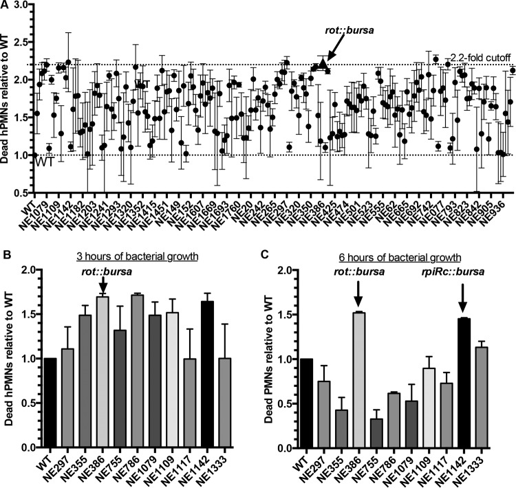
Staphylococcus aureus is a formidable human pathogen that uses secreted cytolytic factors to injure immune cells and promote infection of its host. Of these proteins, the bicomponent family of pore-forming leukocidins play critical roles in S. aureus pathogenesis. The regulatory mechanisms governing the expression of these toxins are incompletely defined. In this work, we performed a screen to identify transcriptional regulators involved in leukocidin expression in S. aureus strain USA300. We discovered that a metabolic sensor-regulator, RpiRc, is a potent and selective repressor of two leukocidins, LukED and LukSF-PV. Whole-genome transcriptomics, S. aureus exoprotein proteomics, and metabolomic analyses revealed that RpiRc influences the expression and production of disparate virulence factors. Additionally, RpiRc altered metabolic fluxes in the trichloroacetic acid cycle, glycolysis, and amino acid metabolism. Using mutational analyses, we confirmed and extended the observation that RpiRc signals through the accessory gene regulatory (Agr) quorum-sensing system in USA300. Specifically, RpiRc represses the rnaIII promoter, resulting in increased repressor of toxins (Rot) levels, which in turn negatively affect leukocidin expression. Inactivation of rpiRc phenocopied rot deletion and increased S. aureus killing of primary human polymorphonuclear leukocytes and the pathogenesis of bloodstream infection in vivo. Collectively, our results suggest that S. aureus senses metabolic shifts by RpiRc to differentially regulate the expression of leukocidins and to promote invasive disease.

Importance: The bicomponent pore-forming leukocidins play pivotal roles in the ability of S. aureus to kill multiple host immune cells, thus enabling this pathogen to have diverse tissue- and species-tropic effects. While the mechanisms of leukocidin-host receptor interactions have been studied in detail, the regulatory aspects of leukocidin expression are less well characterized. Moreover, the expression of the leukocidins is highly modular in vitro, suggesting the presence of regulators other than the known Agr, Rot, and S. aureus exoprotein pathways. Here, we describe how RpiRc, a metabolite-sensing transcription factor, mediates the repression of two specific leukocidin genes, lukED and pvl, which in turn has complex effects on the pathogenesis of S. aureus Our findings highlight the intricacies of leukocidin regulation by S. aureus and demonstrate the involvement of factors beyond traditional virulence factor regulators.


Figures

FIG 1
FIG 1
Leukocidin protein abundances and promoter activities vary during growth in vitro. (A) Quantitative mass spectrometry analyses (by LFQ) of postexponentially grown USA300 JE2 culture filtrates. (B) Leukocidin promoter activity in TSB measured by luminescence of postexponentially grown USA300 harboring plasmids of leukocidin promoter sequences fused to the luciferase gene. The values shown are averages of two independent experiments each performed with three colonies of each strain ± the standard deviation.
FIG 2
FIG 2
Identification of transcriptional regulators that enhance S. aureus cytotoxicity. (A) Primary intoxication screening of hPMNs with USA300 JE2 supernatants at a final concentration of 5% (vol/vol). Data points represent neutrophil death caused by an individual mutant relative to that caused by wild-type (WT) bacteria (lower dotted line). Supernatants from each mutant were tested on hPMNs from four donors, and cell viability was measured with CellTiter metabolic dye. A 2.2-fold cutoff was used to identify candidate mutants for further screening (upper dotted line). The data point indicated by the triangle represents the cytotoxicity of a rot::bursa mutant. (B and C) Validation intoxication screening of hPMNs isolated from two donors with supernatants from select S. aureus mutants. Wild-type and mutant bacteria were grown for 3 (B) and 6 (C) h postinoculation. Error bars indicate the standard error of the mean.
FIG 3
FIG 3
RpiRc is a potent regulator of S. aureus secreted proteins. (A) Exoprotein profiles of USA300 JE2 wild-type (WT) and rpiR mutant bacteria, as assessed by Coomassie staining. The asterisk indicates the approximate leukocidin protein size. (B) Exoprotein profile of USA300 LAC wild-type, rpiRc, and rpiRc+ isogenic strains. (C) Heat map of LAC wild-type and rpiRc mutant secretomes as assessed by mass spectrometry. (D to F) Levels of exoenzymes (D), surface and immunomodulatory proteins (E), and cytotoxins (F) ± the standard deviation in exoproteomes of wild-type versus rpiRc mutant USA300 LAC.
FIG 4
FIG 4
Defining the RpiRc regulon and the RpiRc-associated metabolic changes. RNA-Seq of USA300 wild-type and rpiRc mutant bacteria. (A) Genomic map depicting transcription profiles. The outer circle (blue) indicates the RPKM (number of reads per kilobase per million mapped reads) of the wild type, and the inner circle (red) indicates the RPKM of the rpiRc mutant. The center circle is a heat map showing the fold differences in expression. Genes that were 5-fold upregulated (blue arrows) or 5-fold downregulated (red arrows) are indicated. (B) Fold differences in metabolic flux between the wild type (WT) and the rpiRc mutant based on RNA-Seq results. The enzyme classification (E.C.) numbers represent specific enzymatic reactions and their corresponding pathways. (C) Fold differences in transcript (RNA-Seq) and protein (proteomics) abundance of the individual leukocidins between wild-type and rpiRc mutant bacteria.
FIG 5
FIG 5
RpiRc regulates leukocidin expression by acting on Rot translation. (A) qRT-PCR analyses of transcripts in the USA300 LAC strains indicated. The relative abundance of the individual gene products was normalized to that of the wild type (WT). The experiment was performed with RNA extracted from three individual colonies, each assayed in triplicate, ± the standard deviation. (B) Time course comparing the expression of various promoters fused to luciferase or fluorescence genes. The values are averages of two independent experiments, each performed with three independent colonies of each strain, ± the standard deviation.
FIG 6
FIG 6
Inactivation of rpiRc enhances the virulence of USA300 LAC. (A) Intoxication of hPMNs (from six donors ± the standard of the mean) with a 2-fold titration of USA300 culture filtrates. Cell death was measured with CellTiter metabolic dye. (B) Infection of hPMNs (from three donors ± the standard of the mean) with the isogenic USA300 strains indicated under nonphagocytosing conditions. Cell death was measured as percent lactate dehydrogenase (LDH) release from lysed hPMNs. Statistical analyses were performed by ANOVA with Dunnett’s multiple comparison test. The asterisk indicates a P value of <0.05 for wild-type (WT) versus rpiRc::bursa bacteria and for rpiRc::bursa versus rpiRc::bursa/rpiRc+ bacteria. (C) Growth rebound of the opsonized USA300 isogenic strains indicated during infection of hPMNs (from three donors ± the standard of the mean) at an MOI of 10 under phagocytosing conditions. Percent survival is the CFU count enumerated at the different time points relative to the start of infection. Statistical analyses were performed by ANOVA with Dunnett’s multiple comparison test. (D) Kaplan-Meier curve showing the percent survival of mice (10/group) infected retro-orbitally with ~5 × 10⁷ CFU of the isogenic strains indicated. Statistically significant differences between curves were determined by log rank (Mantel-Cox) test, and P values are shown. The results were corrected for multiple comparisons by using the Bonferroni-corrected threshold (assumed a statistical significance of 0.05 divided by two comparisons; 0.025). (E) Scores of murine skin lesions and dermonecrosis (five mice per group with two abscesses each) 48 h after intradermal infection with ~1 × 10⁶ CFU of the strains indicated. Each symbol is the average score per site of infection. Statistical analyses were performed with the Kruskal-Wallis test. *, P < 0.05; **, P < 0.005. Representative pictures of skin lesions at 48 h postinfection are shown on the right.
FIG 7
FIG 7
Mutation of rpiRc enhances S. aureus Newman virulence in a murine bloodstream infection model through derepression of LukED. (A) At the top is the exoprotein profile of postexponentially grown isogenic Newman strains stained with Coomassie blue (CB). The asterisk indicates the approximate leukocidin size. At the bottom is an immunoblot (IB) detecting LukD in the same samples. (B) Intoxication of hPMNs (from six donors ± the standard error of mean) with a 2-fold titration of Newman culture filtrates. Cell death was measured with CellTiter metabolic dye. (C and D) Bacteria recovered from the livers (C) and hearts (D) of female ND4 Swiss-Webster mice infected retro-orbitally with 1 × 10⁷ CFU of the strains indicated. Each symbol represents a mouse (n = 10). Analyses of statistical significance were performed with the Kruskal-Wallis test. *, P < 0.05; **, P < 0.005; ***, P < 0.0005. (E) Kaplan-Meier curve showing the percent survival of mice (20 per group) infected retro-orbitally with 2.5 × 10⁷ CFU of the isogenic strains indicated. Statistically significant differences between curves were determined by log rank (Mantel-Cox) test, and P values are shown. The results were corrected for multiple comparisons by using the Bonferroni-corrected threshold (assumed statistical significance of 0.05 divided by three comparisons; 0.0167), and the statistical significance of differences was determined. WT, wild type.

References

    1. Vandenesch F, Lina G, Henry T. 2012. Staphylococcus aureus hemolysins, bi-component leukocidins, and cytolytic peptides: a redundant arsenal of membrane-damaging virulence factors? Front Cell Infect Microbiol 2:12. doi:10.3389/fcimb.2012.00012. - DOI - PMC - PubMed
    1. Otto M. 2014. Staphylococcus aureus toxins. Curr Opin Microbiol 17:32–37. doi:10.1016/j.mib.2013.11.004. - DOI - PMC - PubMed
    1. Alonzo F III, Torres VJ. 2014. The bicomponent pore-forming leucocidins of Staphylococcus aureus. Microbiol Mol Biol Rev 78:199–230. doi:10.1128/MMBR.00055-13. - DOI - PMC - PubMed
    1. Menestrina G, Dalla Serra M, Comai M, Coraiola M, Viero G, Werner S, Colin DA, Monteil H, Prévost G. 2003. Ion channels and bacterial infection: the case of beta-barrel pore-forming protein toxins of Staphylococcus aureus. FEBS Lett 552:54–60. doi:10.1016/S0014-5793(03)00850-0. - DOI - PubMed
    1. DuMont AL, Torres VJ. 2014. Cell targeting by the Staphylococcus aureus pore-forming toxins: it’s not just about lipids. Trends Microbiol 22:21–27. doi:10.1016/j.tim.2013.10.004. - DOI - PMC - PubMed

Publication types

MeSH terms

LinkOut - more resources