. 2016 Aug 15;25(16):3407-3415.
doi: 10.1093/hmg/ddw183. Epub 2016 Jun 21.

A novel missense variant in PRKCB segregates low-frequency hearing loss in an autosomal dominant family with Meniere's disease

Affiliations

A novel missense variant in PRKCB segregates low-frequency hearing loss in an autosomal dominant family with Meniere's disease

Carmen Martín-Sierra et al. Hum Mol Genet. .

Abstract

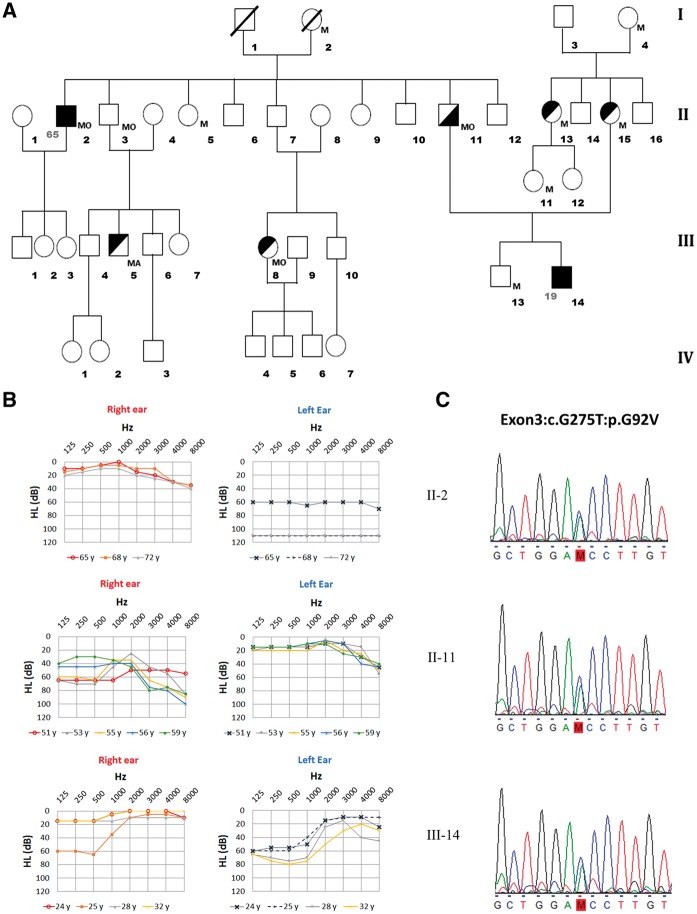
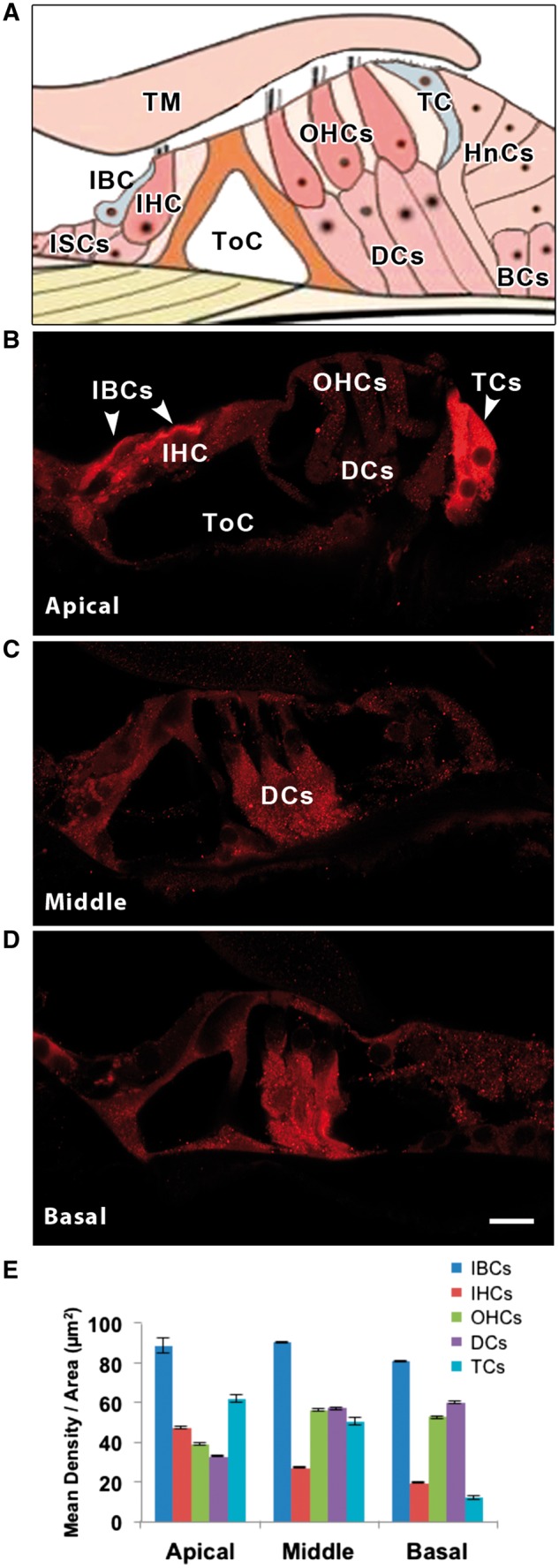
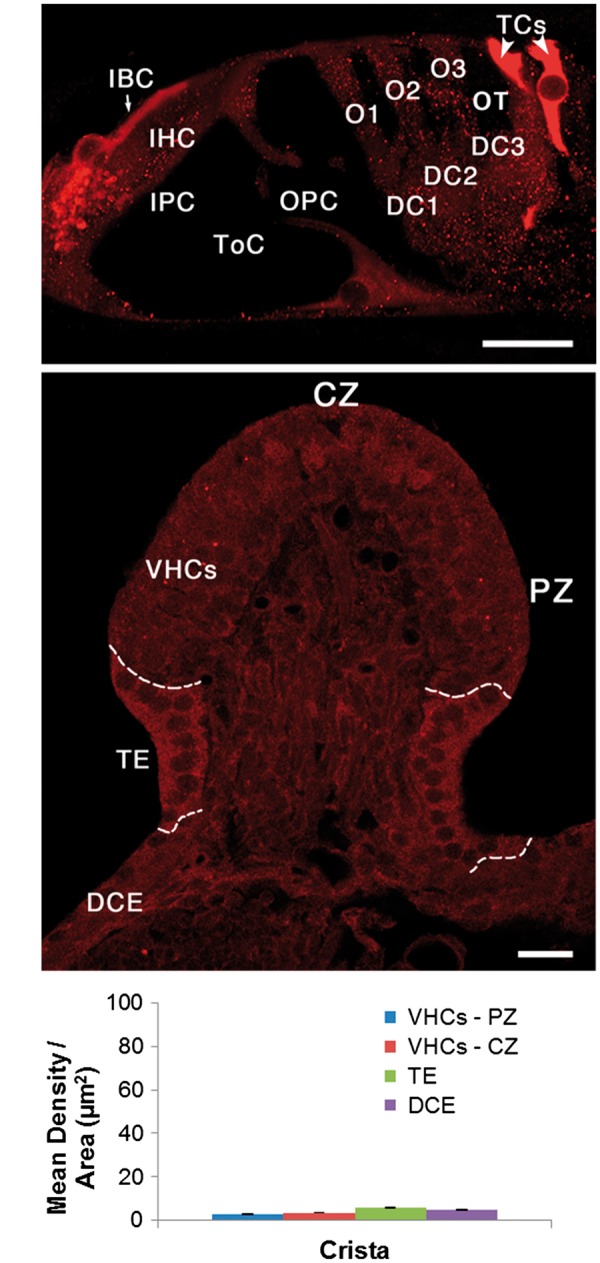
Meniere's Disease (MD) is a complex disorder associated with an accumulation of endolymph in the membranous labyrinth in the inner ear. It is characterized by recurrent attacks of spontaneous vertigo associated with sensorineural hearing loss (SNHL) and tinnitus. The SNHL usually starts at low and medium frequencies with a variable progression to high frequencies. We identified a novel missense variant in the PRKCB gene in a Spanish family with MD segregating low-to-middle frequency SNHL. Confocal imaging showed strong PKCB II protein labelling in non-sensory cells, the tectal cells and inner border cells of the rat organ of Corti with a tonotopic expression gradient. The PKCB II signal was more pronounced in the apical turn of the cochlea when compared with the middle and basal turns. It was also much higher in cochlear tissue than in vestibular tissue. Taken together, our findings identify PRKCB gene as a novel candidate gene for familial MD and its expression gradient in supporting cells of the organ of Corti deserves attention, given the role of supporting cells in K+ recycling within the endolymph, and its apical turn location may explain the onset of hearing loss at low frequencies in MD.


Figures

Figure 1.
Figure 1.
Spanish family with Meniere’s disease with a novel variant in the PRKCB gene. (A) Pedigree of a family presenting AD FMD with three affected men in two generations. Symbols in the pedigree represent: complete MD phenotype (full black square), numbers at the left of the symbol indicate the age of onset; incomplete phenotype (partial black square or circle, upper-left black: vestibular phenotype; lower-right black: cochlear phenotype). Abbreviations: M, Migraine; MA, Migraine with aura; MO, Migraine without aura. (B) Air-conduction audiograms showing a severe sensorineural hearing loss starting at low frequencies in affected individuals at several ages (age symbols shown below the chart); upper X axis represents frequencies in Hz, and Y axis represents hearing level (HL) in dB. (C) Genomic sequence at PRKCB gene (chr16:23999898G > T) with the novel variant (M) segregating hearing loss (indicated in red).
Figure 2.
Figure 2.
(A) Schematic diagram of the organ of Corti to show the location of tectal cells (TCs) and inner border cells (IBCs). PKCB II immuno-labelling in the three turns of the rat cochlea, apical (B), middle (C) and basal (D). Cell types are labelled in A, B and C. Note that the apical IBCs and TCs are most intensely labelled, followed by basal and middle turn Deiter’s cells (DCs). Hair cells, both inner (IHCs) and outer (OHCs), are barely above background level. Other abbreviations: tectorial membrane (TM), inner sulcus cells (ISCs), tunnel of Corti (ToC), Hensen’s cells (HnCs), Boettcher’s cells (BCs). (E) Mean density per area of PKCB II for each cochlear cell type and turn of the cochlea. Scale bar in D= 10 µm, applies to B-D.
Figure 3.
Figure 3.
Here we directly compare cochlear PKCB II immuno-labelling in the apical turn of (A) the rat cochlea with (B) the vestibular crista ampullaris in the same animal. Note that the entire vestibular epithelium is less intensely labelled than the cochlear inner border cells (IBCs) and tectal cells (TCs). Dashed lines in B separate the vestibular sensory epithelium (VHCs) from the transitional epithelium (TE), and the TE from the dark cell epithelium (DCE). (C) Mean density per area of PKCB II in different regions of the rat crista ampullaris. Note this histogram is the same scale as that in Fig. 2E. Other abbreviations: IHC, inner hair cell; IPC, inner pillar cell; ToC, tunnel of Corti; OPC, outer pillar cell; O1, O2, O3, first, second and third row outer hair cells; DC1, DC2, DC3, first, second and third row Deiters cells; OT, outer tunnel. CZ, central zone; PZ, peripheral zone; VHCs, vestibular hair cells (sensory epithelium), TE transitional epithelium (supporting cells). Scale bars = 10 µm.

References

    1. Corti A. (1851) Recherches sur l’organe de l'ouïe des mammiferes. Limaçon. Z. Wiss. Zool, 3, 106–169.
    1. Frucht C.S., Uduman M., Kleinstein S.H., Santos-Sacchi J., Navaratnam D.S. (2011) Gene expression gradients along the tonotopic axis of the chicken auditory epithelium. J. Assoc. Res. Otolaryngol., 12, 423–435. - PMC - PubMed
    1. Yoshimura H., Takumi Y., Nishio S.Y., Suzuki N., Iwasa Y., Usami S. (2014) Deafness gene expression patterns in the mouse cochlea found by microarray analysis. PLoS One., 9, e92547.. - PMC - PubMed
    1. Lopez-Escamez J.A., Carey J., Chung W.H., Goebel J.A., Magnusson M., Mandala M., Newman-Toker D.E., Strupp M., Suzuki M., Trabalzini F., et al. (2015) Diagnostic criteria for Meniere's disease. J. Vestib. Res., 25, 1–7. - PubMed
    1. Harcourt J., Barraclough K., Bronstein A.M. (2014) Meniere's disease. Bmj., 349, g6544.. - PubMed

Publication types