. 2016 Sep:133:118-128.
doi: 10.1016/j.nlm.2016.06.016. Epub 2016 Jun 18.

Temporary inactivation reveals that the CA1 region of the mouse dorsal hippocampus plays an equivalent role in the retrieval of long-term object memory and spatial memory

Affiliations

Temporary inactivation reveals that the CA1 region of the mouse dorsal hippocampus plays an equivalent role in the retrieval of long-term object memory and spatial memory

Robert W Stackman Jr et al. Neurobiol Learn Mem. 2016 Sep.

Abstract

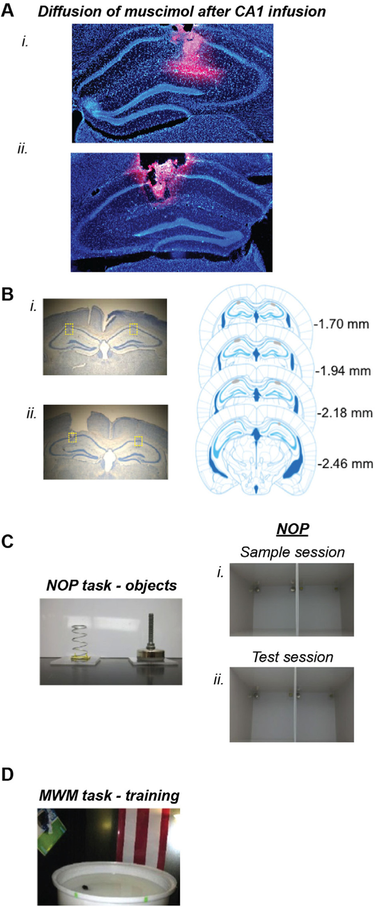
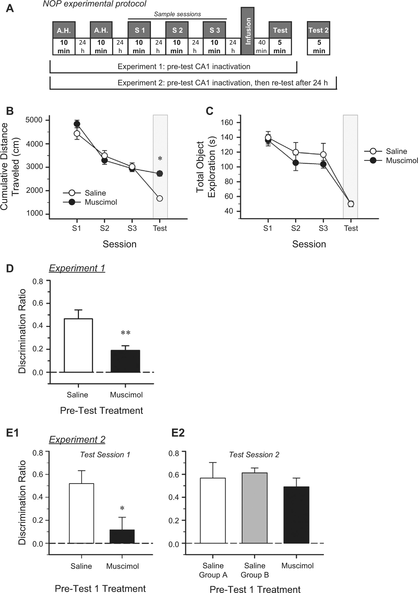
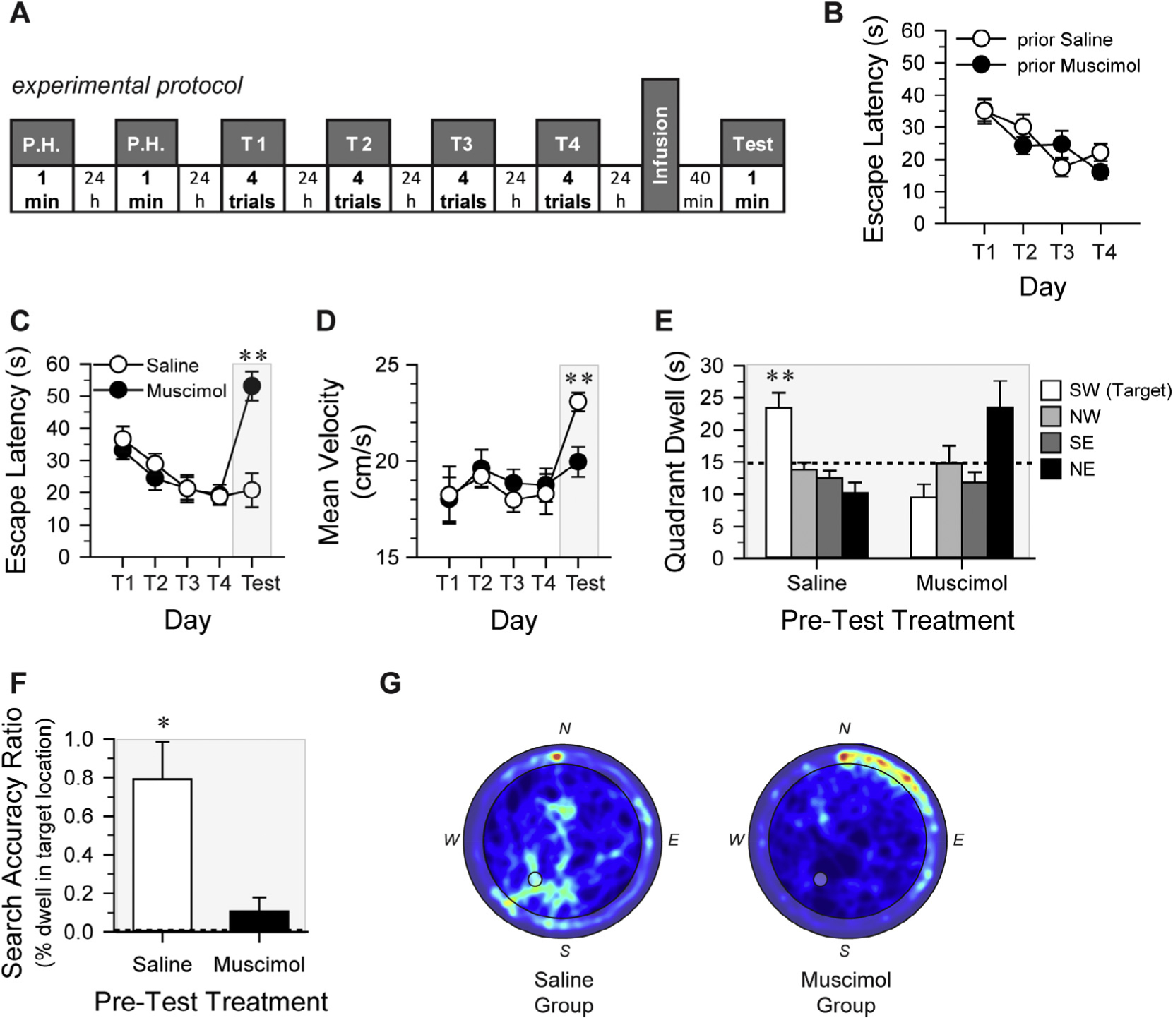
Recognition of a previously experienced item or object depends upon the successful retrieval of memory for the object. The neural mechanisms that support object recognition memory in the mammalian brain are not well understood. The rodent hippocampus plays a well-established role in spatial memory, and we previously demonstrated that temporary inactivation of the mouse hippocampus impairs object memory, as assessed with a novel object preference (NOP) test. The present studies were designed to test some remaining issues regarding the contribution of the CA1 sub-region of the mouse dorsal hippocampus to long-term object memory. Specifically, we examined whether the retrieval of spatial memory (as assessed by the Morris water maze; MWM) and object recognition memory are differentially sensitive to inactivation of the CA1 region. The current study used pre-test local microinfusion of muscimol directly into the CA1 region of dorsal hippocampus to temporarily interrupt its function during the respective retrieval phases of both behavioral tasks, in order to compare the contribution of the CA1 to object memory and spatial memory. Histological analyses revealed that local intra-CA1 injection of muscimol diffused within, and not beyond, the CA1 region of dorsal hippocampus. The degree of memory retrieval impairment induced by muscimol was comparable in the two tasks, supporting the view that object memory and spatial memory depend similarly on the CA1 region of rodent hippocampus. Further, we confirmed that the muscimol-induced impairment of CA1 function is temporary. First, mice that exhibited impaired object memory retrieval immediately after intra-CA1 muscimol, subsequently exhibited unimpaired retrieval of object memory when tested 24h later. Secondly, a cohort of mice that exhibited impaired object memory retrieval after intra-CA1 muscimol later acquired spatial memory in the MWM comparable to that of control mice. Together, these results offer further support for the involvement of the CA1 region of mouse hippocampus in object recognition memory, and provide evidence to suggest that the NOP task is as much a test of hippocampal function as the classic MWM test.

Keywords: Hippocampus; Morris water maze; Muscimol; Novel object preference; Object recognition; Spatial memory.


Figures

Fig. 1.
Fig. 1.
Diffusion of muscimol, histological verification, objects and testing arenas.A. (i and ii), Representative photomicrographs of coronal sections through the dorsal hippocampus from three mice that received bilateral infusions of fluorophore-conjugated muscimol (BODIPY TMR-X, Molecular Probes). Sections were mounted using DAPI fluoromount to improve visualization of the locally infused fluorophore-conjugated muscimol. Images reveal that the red fluorescence (i.e., the fluorescent conjugated muscimol) diffused within the hippocampus, but remained within the CA1 region of the dorsal hippocampus. B. left, Representative photomicrographs of Cresyl violet-stained sections from two mice indicating accurate placement of guide cannulae above the CA1 region of the dorsal hippocampus and the infusion cannulae tracks into the CA1. right, Composite montage depicting the distribution of bilateral microinfusion placements into the CA1 region of the dorsal hippocampus from all mice included in the data analyses from both Experiments 1 and 2. All infusions were confirmed to be within the region depicted by gray. Numbers refer to distance (in mm) from bregma; plates are modified from the respective coronal plates of the Franklin and Paxinos (2008) mouse stereotaxic atlas. C. Left, Photographs of the objects used in the NOP experiments. Right, Photograph depicting the configuration of objects within both arenas during the sample sessions (i), and the test session (ii). These independent dual chamber configurations permitted two mice to be simultaneously tested during each sample or test session. D. Photograph of the MWM room, depicting approximate locations of a subset of the extra-maze cues and the size of pool. In the image, a mouse is standing on the platform submerged within the SW quadrant of the pool.
Fig. 2.
Fig. 2.
Pre-test intra-CA1 muscimol impairs retrieval of long-term object memory. A. Schematic of the experimental protocols used for the NOP task in Experiments 1 and 2. A.H.: Arena Habituation (exposure to empty arena); S1-S3: Sample Sessions (each of three 10-min exposures (1/day) to a pair of identical objects in the familiar arena; the same objects were used for each sample session). B. Cumulative distance traveled decreased significantly across the 10-min sample sessions (P < 0.001) indicating habituation to the arena; however, there was no difference between the future treatment groups (n.s.). Mice that received pre-test intra-CA1 muscimol traveled a significantly greater distance during the 5-min test session than did the pre-test saline mice, possibly due to impaired retrieval of the memory of the arena. C. There was no significant difference between pre-test treatment groups in total time spent exploring objects over the three 10-min sample sessions or during the 5-min test session. Shaded regions in B and C highlight the session conducted under the influence of the microinfusion. D. Mice that received pre-test intra-CA1 saline exhibited a significantly stronger preference for the novel object than did the pre-test muscimol-treated mice. Dashed line indicates chance performance. All error bars indicate ± S.E.M. * = P < 0.01; ** = P < 0.005. E. In Experiment 2, mice received three sample sessions (1/day) as in Experiment 1. Mice received intra-CA1 saline or muscimol before NOP test session 1. E1. Mice that received intra-hippocampal saline prior to NOP Test session 1 exhibited significant discrimination between the familiar and novel objects. However, the mice treated with muscimol were impaired, consistent with results in D. Dashed line indicates chance performance. All error bars indicate ± S.E.M. * = P < 0.03. E2. Mice that received intra-CA1 saline prior to NOP test session 1 were assigned to either saline group A (exposed to Test session 1 and Test session 2), or saline group B (exposed to only Test session 2). During Test session 2, saline group A again demonstrated significant object discrimination and performed comparably to saline group B. Additionally, the muscimol-treated mice that exhibited impaired discrimination during Test session 1, exhibited significant object discrimination during Test session 2, performing similar to both saline groups. Dashed line indicates chance performance. All error bars indicate ± S.E.M.
Fig. 3.
Fig. 3.
Pre-test intra-CA1 muscimol impairs retrieval of long-term spatial memory and memory for task performance. A. Schematic of the experimental protocol used for the MWM task. P.H.: Pool Habituation (exposure to the pool without extra-maze visual cues, platform in center of pool); T1-T4: Training sessions comprising 4 trials/day (spatial training with extra-maze visual cues and platform in SW quadrant); Test: platform-less probe test of spatial memory retention. Mice received intra-CA1 saline or muscimol 10–11 days prior to the onset of MWM training (see Fig. 2D). B. Escape latency across the four training sessions plotted according to the previous intra-CA1 treatment (prior Saline, prior Muscimol). Prior intra-CA1 treatment did not affect spatial training in the MWM, indicating that the effects of muscimol on CA1 were temporary. Treatment assignments for pre-MWM probe test microinfusions were reversed from that of pre-NOP test session. Escape latency (C) and mean velocity (D) plotted according to MWM pre-probe test treatment. There was no effect of future treatment on either dependent measure across training trial blocks. However, pre-test muscimol-treated mice took a significantly longer latency to reach the platform location than did the pre-test saline-treated mice (P < 0.001), indicating impaired retrieval of spatial memory. The mean velocity of pre-test saline-treated mice was significantly greater than that of the pre-test muscimol-treated mice. E. Mice that received pre-probe test intra-CA1 saline spent significantly more time in the target SW quadrant than in any other quadrant, and more time in the target quadrant than did the muscimol-treated mice. Although pre-test intra-CA1 muscimol-treated mice spent more time in the NE quadrant, they failed to exhibit a statistically significant preference for any quadrant. Dashed line indicates chance performance: 15 s dwell in a given quadrant. F. Specific search accuracy was calculated by finding the percentage of time during the 60-s probe test that each mouse spent in the exact location of the pool where the platform had been during training. Pre-test saline-treated mice exhibited a significantly greater search accuracy ratio than would be expected by chance (see dashed line at 0.005696; P = 0.002); however, muscimol-treated mice did not perform better than chance. G. Composite map of all swim paths during the probe test for each pre-test treatment condition. Plots present quadrant dwell as a “heat map” representing mean dwell times according to a color code with darker, cooler colors indicating low dwell times and warmer colors indicating high dwell times. The pre-test saline-treated mice appropriately concentrated their search in the SW quadrant, while the pre-test muscimol-treated mice failed to concentrate their search in the SW quadrant and instead demonstrate predominant thigmotaxis. The gray circle in center of the SW quadrant of each composite image indicates the location where the platform had been submerged during training. All error bars indicate ± S.E.M. *, P < 0.01; **, P ≤ 0.001. The gray shaded boxes in plots C–F indicate behavioral testing that occurred under the influence of the respective intra-CA1 treatment.
Fig. 4.
Fig. 4.
Pre-test inactivation of the CA1 region impairs retrieval of object memory and spatial memory equivalently. To compare the degree of memory impairment produced by intra-CA1 muscimol in the two distinct tasks, discrimination ratio (DR) scores from the NOP experiment and MDtP scores from the MWM experiment were normalized to the best actual score for each experiment. The scatter plot depicts the distribution of scores of individual mice for the respective task according to the intra-CA1 treatment condition; the treatment mean ± SEM normalized scores are also indicated (open circles, saline; filled squares, muscimol). Note: overlap in data points prevents all individual scores from being visible. In addition to being significantly different from the respective muscimol condition, the scores of the saline-treated mice were consistent across the two experiments (all comparisons, P > 0.05). The scores of the muscimol-treated mice were also consistent across the two experiments (all comparisons, P > 0.05), indicating that intra-CA1 muscimol impaired object memory and spatial memory retrieval to a comparable degree. All error bars indicate ± SEM.

References

    1. Baker KB, & Kim JJ (2002). Effects of stress and hippocampal NMDA receptor antagonism on recognition memory in rats. Learning & Memory, 9, 58–65. - PMC - PubMed
    1. Barker GR, & Warburton EC (2011). When is the hippocampus involved in recognition memory? Journal of Neuroscience, 31, 10721–10731. - PMC - PubMed
    1. Barker GR, Warburton EC, Koder T, Dolman NP, More JC, Aggleton JP, … Brown MW (2006). The different effects on recognition memory of perirhinal kainate and NMDA glutamate receptor antagonism: Implications for underlying plasticity mechanisms. The Journal of Neuroscience: The Official Journal of the Society for Neuroscience, 26, 3561–3566. - PMC - PubMed
    1. Beer Z, Chwiesko C, Kitsukawa T, & Sauvage MM (2013). Spatial and stimulus-type tuning in the LEC, MEC, POR, PrC, CA1, and CA3 during spontaneous item recognition memory. Hippocampus, 23, 1425–1438. - PubMed
    1. Berlyne DE (1950). Novelty and curiosity as determinants of exploratory behaviour. British Journal of Psychology. General Section, 41, 68–80.

MeSH terms