Review
. 2016 Dec;57(12):2099-2114.
doi: 10.1194/jlr.R066514. Epub 2016 Jun 21.

Lipid signaling and lipotoxicity in metaflammation: indications for metabolic disease pathogenesis and treatment

Affiliations
Review

Lipid signaling and lipotoxicity in metaflammation: indications for metabolic disease pathogenesis and treatment

Meric Erikci Ertunc et al. J Lipid Res. 2016 Dec.

Abstract

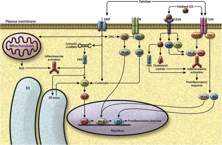
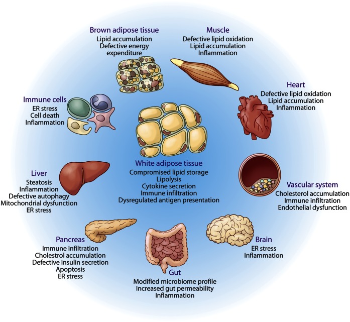
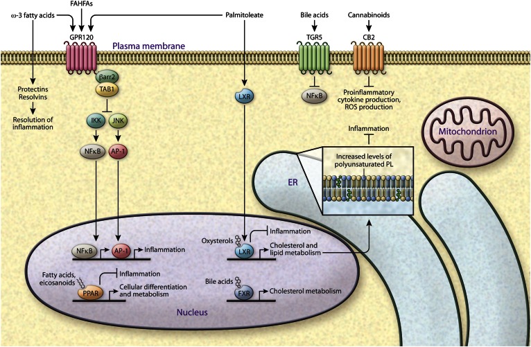
Lipids encompass a wide variety of molecules such as fatty acids, sterols, phospholipids, and triglycerides. These molecules represent a highly efficient energy resource and can act as structural elements of membranes or as signaling molecules that regulate metabolic homeostasis through many mechanisms. Cells possess an integrated set of response systems to adapt to stresses such as those imposed by nutrient fluctuations during feeding-fasting cycles. While lipids are pivotal for these homeostatic processes, they can also contribute to detrimental metabolic outcomes. When metabolic stress becomes chronic and adaptive mechanisms are overwhelmed, as occurs during prolonged nutrient excess or obesity, lipid influx can exceed the adipose tissue storage capacity and result in accumulation of harmful lipid species at ectopic sites such as liver and muscle. As lipid metabolism and immune responses are highly integrated, accumulation of harmful lipids or generation of signaling intermediates can interfere with immune regulation in multiple tissues, causing a vicious cycle of immune-metabolic dysregulation. In this review, we summarize the role of lipotoxicity in metaflammation at the molecular and tissue level, describe the significance of anti-inflammatory lipids in metabolic homeostasis, and discuss the potential of therapeutic approaches targeting pathways at the intersection of lipid metabolism and immune function.

Keywords: diabetes; inflammation; insulin resistance; lipids; obesity; signaling lipids.


Figures

Fig. 1.
Fig. 1.
Coupling of toxic and pro-inflammatory lipids and innate immune response. Accumulation of toxic lipid classes causes deterioration of metabolic regulation, and the effects of such lipids converge on inflammatory and stress pathways. Saturated fatty acids such as palmitate have been extensively studied for their effects in increasing inflammation and inhibiting insulin action. Palmitate is taken up by the cells via FATPs, and is involved in upregulation of cytosolic snoRNAs, which are implicated in ER stress. PKR is a potential kinase that links palmitate-mediated snoRNA upregulation to ER stress induction. PKR also activates inflammasomes and JNK, and promotes AP-1-mediated inflammatory gene expression. Lipotoxicity can also lead to ROS production from mitochondria, which is linked to inflammasome activation. Palmitate directly contributes to the synthesis of DAGs and ceramides. DAGs activate stress kinases, PKCs, and the NFκB pathway; ceramides activate JNK signaling; and both DAGs and ceramides cause insulin resistance via inhibition of IRS1 and AKT, respectively, downstream of insulin receptor (IR). Palmitate can also activate TLR4 signaling, which leads to activation of inflammasomes and induction of inflammatory gene transcription factors interferon regulatory factor (IRF), NFκB, and AP-1. Cholesterol is taken up by the cells via scavenger receptors (e.g., CD36). Accumulation of oxidized cholesterol or cholesterol crystals also leads to induction of TLR4, PKR, and stress kinase (JNK and p38) signaling or inflammasome activation and pro-inflammatory gene expression, which are central players in atherosclerosis progression.
Fig. 2.
Fig. 2.
Integrated organ pathology resulting from lipotoxicity and metabolic inflammation. White adipose tissue is the designated lipid storage depot of higher organisms. In the presence of prolonged nutrient excess or metabolic disturbances, the storage capacity of white adipose tissue is exhausted, leading to ectopic deposition of lipids and lipotoxicity in several organs, such as muscle, liver, pancreas, and heart, as depicted and explained in detail in the text. Because the lipid homeostatic pathways converge with stress and immune responses, such responses in affected tissue systems are activated by harmful lipid species. The outcomes of lipotoxicity differ in the various target tissues, for example NASH in liver, cardiac failure in the heart, altered feeding behavior and appetite due to lipotoxicity in brain, degenerative changes in muscle and BAT, etc. Furthermore, lipotoxicity leads to sustained and unresolved inflammation, organelle dysfunction and stress, which can lead to a vicious cycle of metabolic deterioration.
Fig. 3.
Fig. 3.
Immunometabolic signaling capacity of lipids. The discovery of beneficial roles for specific lipids has changed the perspective on metabolic disease from being excessive fat storage disease to dysregulation of fat composition, and suggested the presence of more complex regulation of lipid subclasses in maintaining homeostatic signaling. The figure depicts several lipids that have anti-inflammatory signaling properties through various distinct mechanisms. For example, lipid ligands bind a variety of nuclear receptors: PPAR ligands are fatty acyl derivatives, LXR ligands are oxysterols, and FXR ligands are bile acids, all with ability to activate an anti-inflammatory program. Lipids are also involved in cell-to-cell and inter-organ communication. Palmitoleate, FAHFAs, and ω-3 fatty acids can activate GPR120 signaling leading to inhibition of JNK- and IKK-mediated inflammation. FAHFAs and palmitoleate are endogenous lipids identified in mouse models of increased adipose tissue de novo lipogenesis, while ω-3 fatty acids are acquired from food intake. The ω-3 fatty acids can further be metabolized into resolvins and protectins, which are involved in resolution of inflammation. Bile acids and endocannabinoids bind to their respective receptors, TGR5 and CB2, to inhibit inflammation and can impact metabolic homeostasis.

References

    1. Hotamisligil G. S. 2006. Inflammation and metabolic disorders. Nature. 444: 860–867. - PubMed
    1. Fu S., Watkins S. M., and Hotamisligil G. S.. 2012. The role of endoplasmic reticulum in hepatic lipid homeostasis and stress signaling. Cell Metab. 15: 623–634. - PubMed
    1. Chiu H. C., Kovacs A., Blanton R. M., Han X., Courtois M., Weinheimer C. J., Yamada K. A., Brunet S., Xu H., Nerbonne J. M., et al. 2005. Transgenic expression of fatty acid transport protein 1 in the heart causes lipotoxic cardiomyopathy. Circ. Res. 96: 225–233. - PubMed
    1. Fu S., Yang L., Li P., Hofmann O., Dicker L., Hide W., Lin X., Watkins S. M., Ivanov A. R., and Hotamisligil G. S.. 2011. Aberrant lipid metabolism disrupts calcium homeostasis causing liver endoplasmic reticulum stress in obesity. Nature. 473: 528–531. - PMC - PubMed
    1. Fu S., Fan J., Blanco J., Gimenez-Cassina A., Danial N. N., Watkins S. M., and Hotamisligil G. S.. 2012. Polysome profiling in liver identifies dynamic regulation of endoplasmic reticulum translatome by obesity and fasting. PLoS Genet. 8: e1002902. - PMC - PubMed

Publication types

Substances