. 2016 Jul 5;113(27):7527-32.
doi: 10.1073/pnas.1604790113. Epub 2016 Jun 21.

Structures of the orthosomycin antibiotics avilamycin and evernimicin in complex with the bacterial 70S ribosome

Affiliations

Structures of the orthosomycin antibiotics avilamycin and evernimicin in complex with the bacterial 70S ribosome

Stefan Arenz et al. Proc Natl Acad Sci U S A. .

Abstract

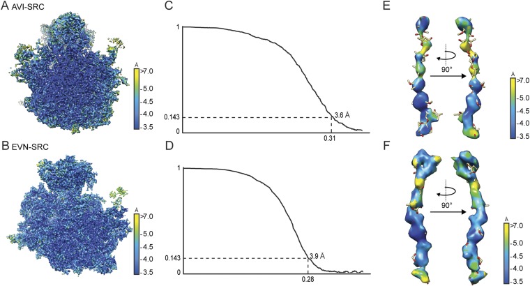
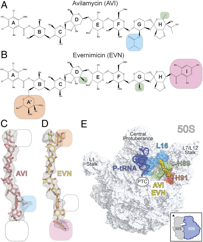
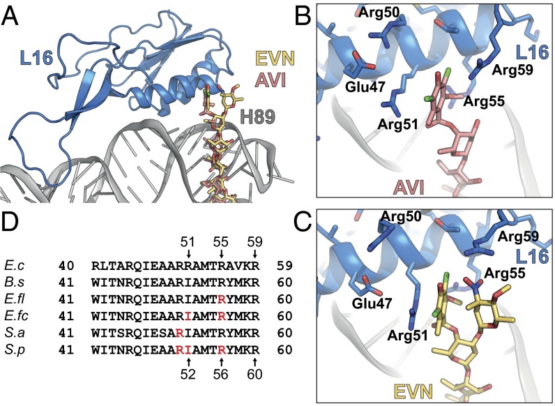
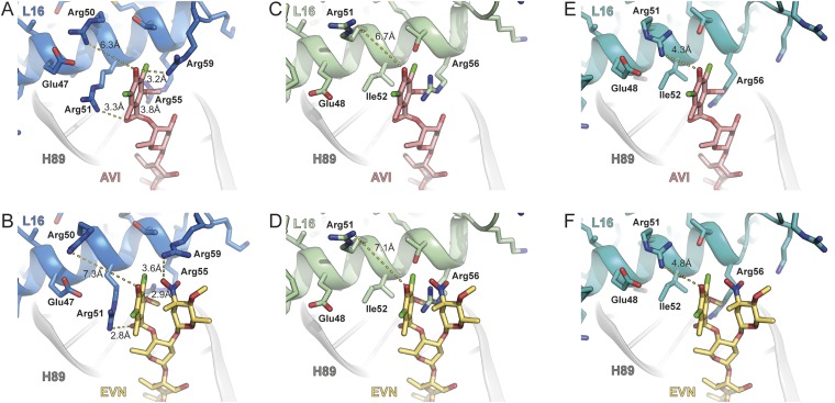
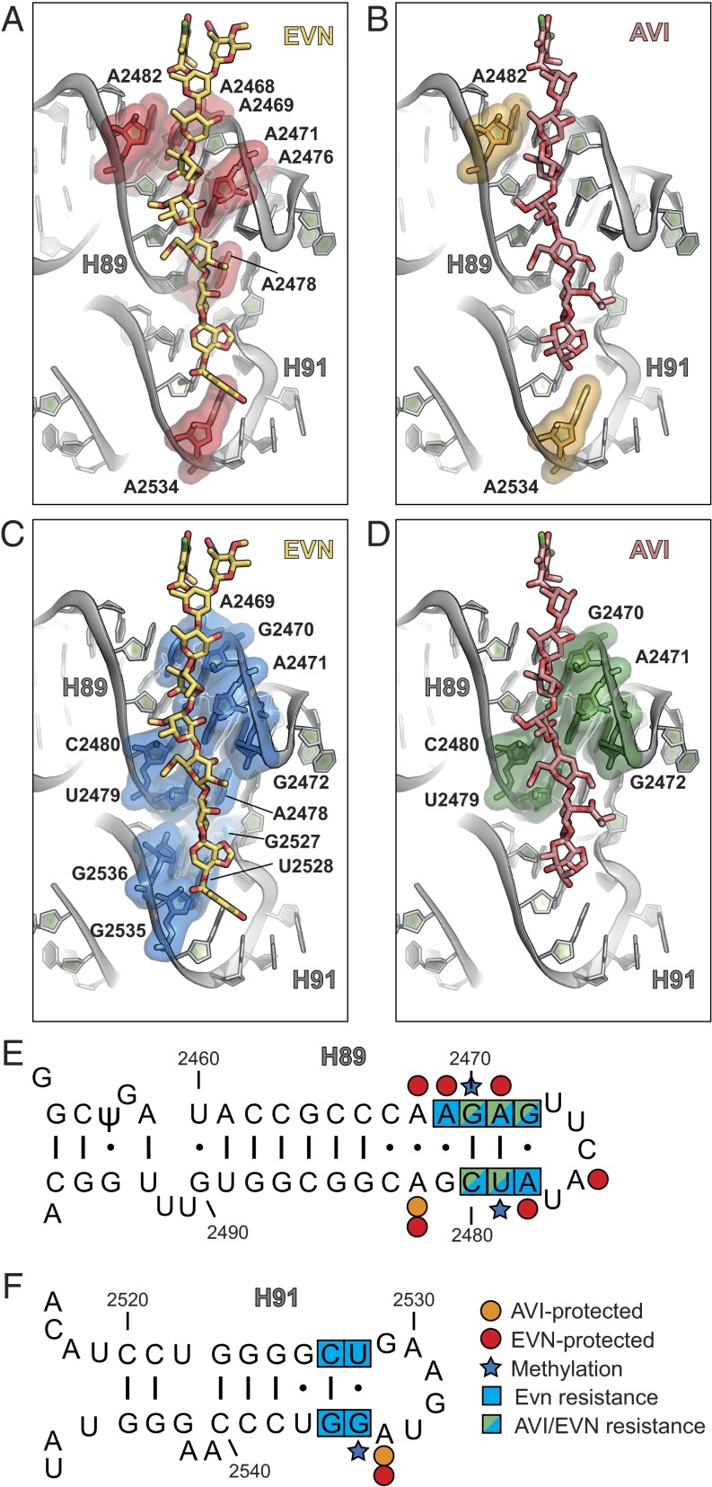
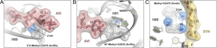
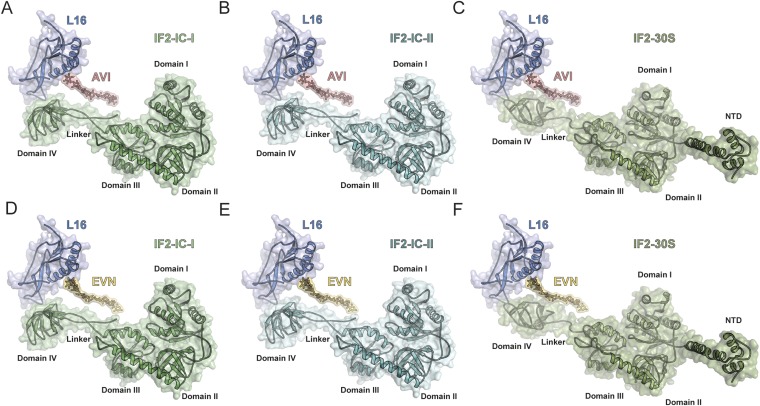
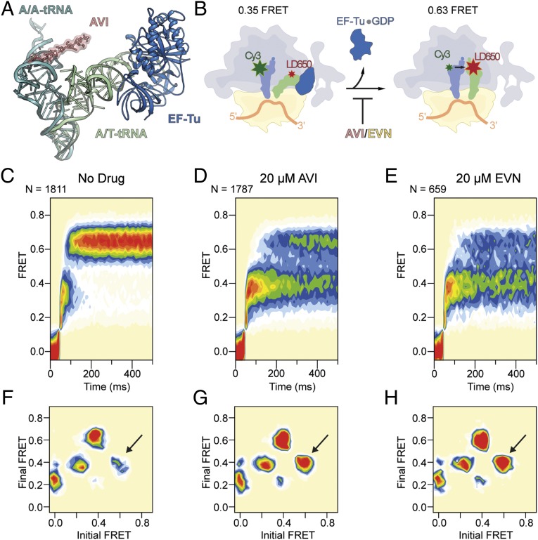
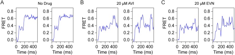
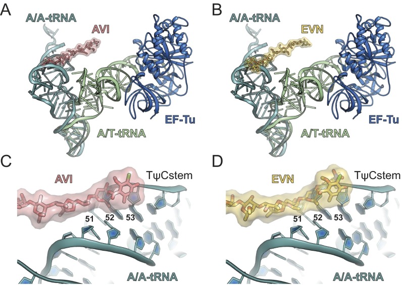
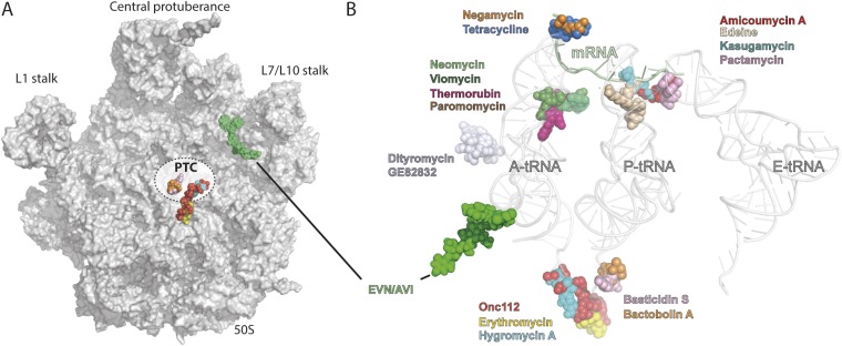
The ribosome is one of the major targets for therapeutic antibiotics; however, the rise in multidrug resistance is a growing threat to the utility of our current arsenal. The orthosomycin antibiotics evernimicin (EVN) and avilamycin (AVI) target the ribosome and do not display cross-resistance with any other classes of antibiotics, suggesting that they bind to a unique site on the ribosome and may therefore represent an avenue for development of new antimicrobial agents. Here we present cryo-EM structures of EVN and AVI in complex with the Escherichia coli ribosome at 3.6- to 3.9-Å resolution. The structures reveal that EVN and AVI bind to a single site on the large subunit that is distinct from other known antibiotic binding sites on the ribosome. Both antibiotics adopt an extended conformation spanning the minor grooves of helices 89 and 91 of the 23S rRNA and interacting with arginine residues of ribosomal protein L16. This binding site overlaps with the elbow region of A-site bound tRNA. Consistent with this finding, single-molecule FRET (smFRET) experiments show that both antibiotics interfere with late steps in the accommodation process, wherein aminoacyl-tRNA enters the peptidyltransferase center of the large ribosomal subunit. These data provide a structural and mechanistic rationale for how these antibiotics inhibit the elongation phase of protein synthesis.

Keywords: Ziracin; antimicrobial; cryo-EM; everninomicin; rRNA.


Conflict of interest statement

The authors declare no conflict of interest.

Figures

Fig. S1.
Fig. S1.
Average and local resolution determination of AVI- and EVN-SRCs. (A and B) Transverse section of the cryo-EM reconstructions of the AVI-SRC (A) and the EVN-SRC (B) colored according to local resolution. (C and D) Average resolution of the AVI-SRC (C) and EVN-SRC (D) was 3.6 and 3.9 Å using the Fourier shell correlation (FSC) cutoff value of 0.143. Because of image processing with an absence of spatial frequencies >8 Å, the FSC value of 0.143 was used for average resolution determination (47). (E and F) Local resolution of the density for AVI (E) and EVN (F).
Fig. 1.
Fig. 1.
Cryo-EM reconstructions of EVN- and AVI-SRC. (A and B) Chemical structures of the orthosomycins AVI (A) and EVN (B), with compositional differences highlighted. (C and D) Cryo-EM electron densities (gray mesh) with fitted models for AVI (red; C) and EVN (yellow; D). (E) Overview of EVN/AVI binding site on the 70S ribosome (50S, gray, and 30S subunit omitted for clarity). Binding position of EVN/AVI (yellow) is shown relative to the P-site tRNA (blue), ribosomal protein L16 (cyan), H89 (green), and H91 (red).
Fig. 2.
Fig. 2.
Interactions of EVN and AVI with the ribosomal protein L16. (A) Overview of L16 (blue) interactions with EVN (gold) and AVI (red). (B and C) Close-up views of showing interactions between Arg-51, -55, and -59 of L16 (blue) and ring A of AVI (B) and rings A and A′ of EVN (C). (D) Sequence alignment of the L16 from E. coli (E.c), B. subtilis (B.s), E. faecalis (E.fl), E. faecium (E.fc), S. aureus (S.a), and S. pneumoniae (S.p), with residues conferring resistance to EVN and AVI highlighted in red.
Fig. S2.
Fig. S2.
Comparison of AVI/EVN binding site with respect to E. coli, B. subtilis, and S. aureus L16. Interaction of AVI (red; A) and EVN (gold; B) with E. coli L16 (blue) compared with the relative position of B. subtilis L16 (green; C and D) (29) and S. aureus L16 (cyan; E and F) (30). In E. coli, Arg-50 is 6.3–7.3 Å from ring A of AVI/EVN, whereas the equivalent residue Arg-51 is 6.7–7.1 Å and 4.3–4.8 Å when superimposing AVI/EVN with B. subtilis L16 (green; C and D) (29) and S. aureus L16 (cyan; E and F) (30).
Fig. 3.
Fig. 3.
Interactions of EVN and AVI with H89 and H91 of the 23S rRNA. (A and B) Binding site of EVN (gold) (A) and AVI (red) (B), with nucleotides in H89 and H91 protected from DMS modification highlighted in red and orange, respectively. (C and D) Binding site of EVN (gold) (C) and AVI (red) (D), with resistance mutations in H89 and H91 highlighted in blue and green, respectively. (E and F) Secondary structure of 23S rRNA with zoom on H89 (E) and H91 (F), with nucleotides protected by EVN (red) and AVI (orange), EVN (blue) and AVI (green) resistance mutations and methylations (blue star) as indicated (–18).
Fig. 4.
Fig. 4.
Structural basis for EVN/AVI resistance via methylation of the 23S rRNA residues. (A) The 2′O-methylation of U2479 in H89 by AviRb (14) clashes with the ring F of the drug. (B) N7 methylation of G2535 in H91 by AviRa (14) is located distal from the AVI binding site. (C) Methylation of the 2′OH of the ribose or N2 position in the nucleobase of G2470 by EmtA (13) clashes with EVN (gold), whereas the N7 position is distal to the drug-binding site.
Fig. S3.
Fig. S3.
Comparison of AVI/EVN binding site with respect IF2. Binding position of AVI (red; AC) and EVN (DF) on the E. coli 70S ribosome relative to IF2-IC conformation I (green; A and D) and IF2-IC conformation II (teal; B and E) on the 70S ribosome (33) and IF2-GTP conformation (olive; C and F) on the 30S subunit (34, 35).
Fig. 5.
Fig. 5.
EVN/AVI inhibit accommodation of tRNA into the A site. (A) Comparison of the relative binding positions on the ribosome of AVI (red), EF-Tu (blue), A/T-tRNA (green) (36, 56), and A/A-tRNA (teal) (38). (B) Schematic diagram of smFRET measurements of tRNA selection. After delivery of EF-Tu⋅GTP⋅tRNA ternary complex containing cognate Phe-tRNAPhe(LD650) to the A site of E. coli 70S ribosomes containing tRNAiMet(Cy3) in the P site, tRNA motion can be tracked through the progression of FRET efficiencies from low (0.2) to intermediate (0.35) FRET during initial steps of selection to high (0.63) FRET upon A-site tRNA accommodation, which is inhibited by AVI/EVN. (CE) Ensemble smFRET histograms showing the time course of aa-tRNA selection, imaged in the absence of drugs (C) or in the presence of 20 μM AVI (D) or 20 μM EVN (E). The histograms were postsynchronized by aligning each observed event to the first appearance of nonzero FRET states. (FH) Transition density plots for the data shown in CE, respectively. These 2D histograms juxtapose the FRET efficiencies immediately before and after FRET transitions. As indicated by arrows, EVN and AVI promote reversible transitions between high and intermediate FRET.
Fig. S4.
Fig. S4.
EVN and AVI specifically block accommodation of A-tRNA into the classical state. Representative smFRET traces exemplifying the processes shown in Fig. 5 CE are shown. (A) In the absence of drug, aa-tRNA rapidly achieves the fully accommodated A/A state (0.63 FRET) via reversible forward-sampling from the 0.35 FRET A/T state. (B and C) AVI (B) and EVN (C) inhibit formation of the A/A state, leading to repeated, unsuccessful sampling events.
Fig. S5.
Fig. S5.
Relative position of EVN/AVI to A/T- and A/A-tRNA. (A and B) Comparison of the relative binding position on the ribosome of AVI (red; A) and EVN (B), with EF-Tu (blue) and A/T-tRNA (green) (36, 56), as well as with accommodated A/A-tRNA (teal) (38). (C and D) Zoom showing the overlap between AVI (red; C) and EVN (gold; D) with nucleotides 51–53 with the TΨC stem of the an accommodated A/A-tRNA (teal) (38).
Fig. S6.
Fig. S6.
EVN and AVI block ribosomes at the start codon of the mRNA. Toe-printing assay monitoring translation in the presence of increasing concentrations (1, 10, and 100 μM) of EVN or AVI is shown. Additionally, control reactions without antibiotic (−) or including thiostrepton (Ths, 100 μM) or edeine (Ede, 50 μM) are shown. AUG designates location of the ribosomes stalled at the start codon. C, U, A, and G indicate the sequencing lanes.
Fig. S7.
Fig. S7.
Relative position of EVN/AVI to other ribosome-targeting antibiotics. (A) Overview of ribosomal 50S subunit (gray) with relative position of EVN/AVI (yellow) compared with known antibiotics that target the PTC. (B) Relative position of EVN/AVI (green) compared with known antibiotics that target the small and large ribosomal subunit, with A-, P-, and E-tRNAs shown for reference.

Similar articles

Cited by

References

    1. Wilson DN. Ribosome-targeting antibiotics and mechanisms of bacterial resistance. Nat Rev Microbiol. 2014;12(1):35–48. - PubMed
    1. Wilson DN. The A-Z of bacterial translation inhibitors. Crit Rev Biochem Mol Biol. 2009;44(6):393–433. - PubMed
    1. Wright D. The orthosomycins, a new family of antibiotics. Tetrahedron. 1979;35(10):1209–1327.
    1. Buzzetti F, et al. [Avilamycin] Experientia. 1968;24(4):320–323. - PubMed
    1. Wagman GH, Luedemann GM, Weinstein MJ. Fermentation and isolation of everninomicin. Antimicrob Agents Chemother (Bethesda) 1964;10:33–37. - PubMed

Publication types

MeSH terms