

Magnetic resonance imaging of ultrasmall superparamagnetic iron oxide-labeled exosomes from stem cells: a new method to obtain labeled exosomes
- PMID: 27330291
- PMCID: PMC4898039
- DOI: 10.2147/IJN.S104152
Magnetic resonance imaging of ultrasmall superparamagnetic iron oxide-labeled exosomes from stem cells: a new method to obtain labeled exosomes
Abstract
Purpose: Recent findings indicate that the beneficial effects of adipose stem cells (ASCs), reported in several neurodegenerative experimental models, could be due to their paracrine activity mediated by the release of exosomes. The aim of this study was the development and validation of an innovative exosome-labeling protocol that allows to visualize them with magnetic resonance imaging (MRI).
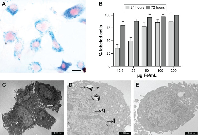
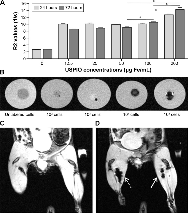
Materials and methods: At first, ASCs were labeled using ultrasmall superparamagnetic iron oxide nanoparticles (USPIO, 4-6 nm), and optimal parameters to label ASCs in terms of cell viability, labeling efficiency, iron content, and magnetic resonance (MR) image contrast were investigated. Exosomes were then isolated from labeled ASCs using a standard isolation protocol. The efficiency of exosome labeling was assessed by acquiring MR images in vitro and in vivo as well as by determining their iron content. Transmission electron microscopy images and histological analysis were performed to validate the results obtained.
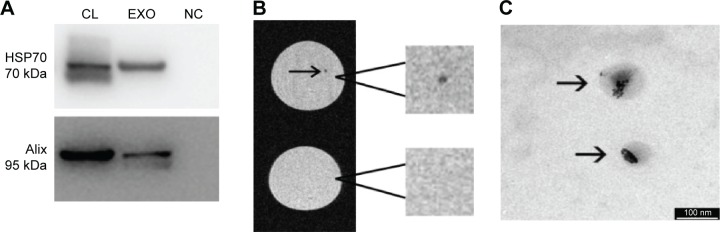
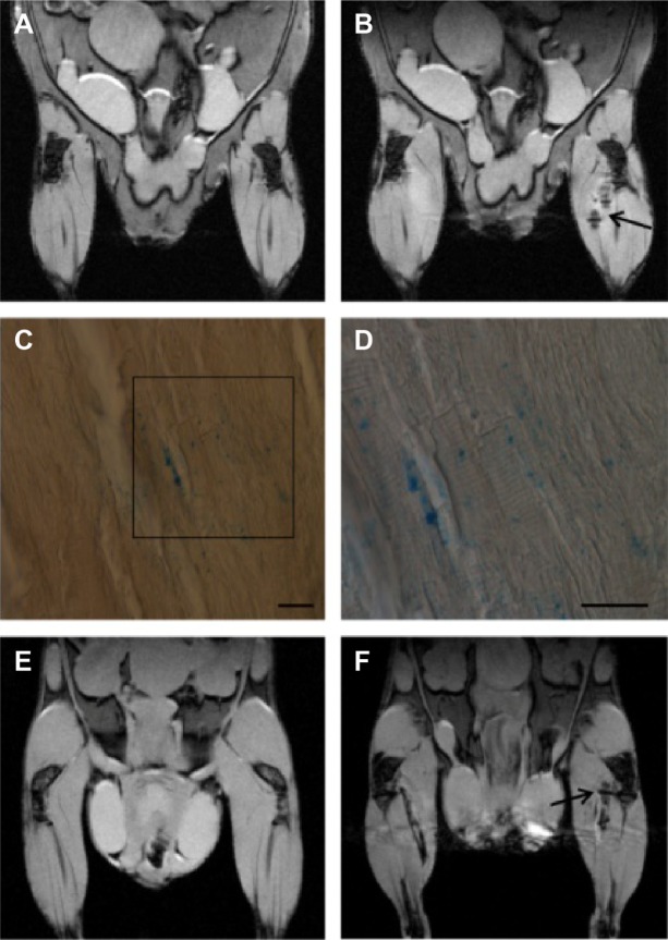
Results: By using optimized experimental parameters for ASC labeling (200 µg Fe/mL of USPIO and 72 hours of incubation), it was possible to label 100% of the cells, while their viability remained comparable to unlabeled cells; the detection limit of MR images was of 10(2) and 2.5×10(3) ASCs in vitro and in vivo, respectively. Exosomes isolated from previously labeled ASCs retain nanoparticles, as demonstrated by transmission electron microscopy images. The detection limit by MRI was 3 µg and 5 µg of exosomes in vitro and in vivo, respectively.
Conclusion: We report a new approach for labeling of exosomes by USPIO that allows detection by MRI while preserving their morphology and physiological characteristics.
Keywords: MRI; cellular imaging; exosome labeling; stem cells labeling; superparamagnetic iron oxide nanoparticles.
Figures
Similar articles
-
Labeling and Magnetic Resonance Imaging of Exosomes Isolated from Adipose Stem Cells.Curr Protoc Cell Biol. 2017 Jun 19;75:3.44.1-3.44.15. doi: 10.1002/cpcb.23. Curr Protoc Cell Biol. 2017. PMID: 28627754
-
Superparamagnetic iron oxide does not affect the viability and function of adipose-derived stem cells, and superparamagnetic iron oxide-enhanced magnetic resonance imaging identifies viable cells.Magn Reson Imaging. 2009 Jan;27(1):108-19. doi: 10.1016/j.mri.2008.05.013. Epub 2008 Jul 26. Magn Reson Imaging. 2009. PMID: 18657922
-
Bioengineered bladder patches constructed from multilayered adipose-derived stem cell sheets for bladder regeneration.Acta Biomater. 2019 Feb;85:131-141. doi: 10.1016/j.actbio.2018.12.016. Epub 2018 Dec 12. Acta Biomater. 2019. PMID: 30553012
-
Methods for magnetically labeling stem and other cells for detection by in vivo magnetic resonance imaging.Cytotherapy. 2004;6(6):621-5. doi: 10.1080/14653240410005267-1. Cytotherapy. 2004. PMID: 15773025 Review.
-
Tracking stem cells with superparamagnetic iron oxide nanoparticles: perspectives and considerations.Int J Nanomedicine. 2017 Jan 25;12:779-793. doi: 10.2147/IJN.S126530. eCollection 2017. Int J Nanomedicine. 2017. PMID: 28182122 Free PMC article. Review.
Cited by
-
Perivascular cell-derived extracellular vesicles stimulate colorectal cancer revascularization after withdrawal of antiangiogenic drugs.J Extracell Vesicles. 2021 May;10(7):e12096. doi: 10.1002/jev2.12096. Epub 2021 May 21. J Extracell Vesicles. 2021. PMID: 34035882 Free PMC article.
-
Engineered exosomes for targeted co-delivery of miR-21 inhibitor and chemotherapeutics to reverse drug resistance in colon cancer.J Nanobiotechnology. 2020 Jan 9;18(1):10. doi: 10.1186/s12951-019-0563-2. J Nanobiotechnology. 2020. PMID: 31918721 Free PMC article.
-
Nanovesicles from adipose-derived mesenchymal stem cells inhibit T lymphocyte trafficking and ameliorate chronic experimental autoimmune encephalomyelitis.Sci Rep. 2018 May 10;8(1):7473. doi: 10.1038/s41598-018-25676-2. Sci Rep. 2018. PMID: 29748664 Free PMC article.
-
Imaging of extracellular vesicles derived from human bone marrow mesenchymal stem cells using fluorescent and magnetic labels.Int J Nanomedicine. 2018 Mar 19;13:1653-1664. doi: 10.2147/IJN.S159404. eCollection 2018. Int J Nanomedicine. 2018. PMID: 29593411 Free PMC article.
-
Exosomes and Extracellular Vesicles as Emerging Theranostic Platforms in Cancer Research.Cells. 2020 Dec 1;9(12):2569. doi: 10.3390/cells9122569. Cells. 2020. PMID: 33271820 Free PMC article. Review.
References
-
- Peroni D, Scambi I, Pasini A, et al. Stem molecular signature of adipose-derived stromal cells. Exp Cell Res. 2008;314(3):603–615. - PubMed
-
- Constantin G, Marconi S, Rossi B, et al. Adipose-derived mesenchymal stem cells ameliorate chronic experimental autoimmune encephalomyelitis. Stem Cell. 2009;27(10):2624–2635. - PubMed
-
- Marconi S, Castiglione G, Turano E, et al. Human adipose-derived mesenchymal stem cells systemically injected promote peripheral nerve regeneration in the mouse model of sciatic crush. Tissue Eng Part A. 2012;18(11–12):1264–1272. - PubMed
MeSH terms
Substances
LinkOut - more resources
Full Text Sources
Other Literature Sources
Medical
Miscellaneous
