

Morphometric changes of corneal endothelial cells following intracameral air for micro perforation of the Descemet Membrane during big-bubble deep anterior lamellar keratoplasty
- PMID: 27330384
- PMCID: PMC4908048
- DOI: 10.1016/j.sjopt.2016.01.003
Morphometric changes of corneal endothelial cells following intracameral air for micro perforation of the Descemet Membrane during big-bubble deep anterior lamellar keratoplasty
Abstract
Aim: The aim of this study was to assess the effect of intracameral air on the endothelial cell morphometrics.
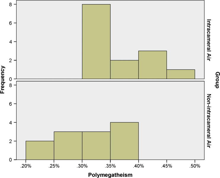
Patients and methods: This is a retrospective controlled interventional cohort study of 26 patients (18 males and 8 females) who underwent unilateral deep anterior lamellar keratoplasty (DALK) for moderate keratoconus. The DALK patients were divided into two groups: a treatment group (14), which had micro perforations of the Descemet Membrane (DM) intraoperatively and received intracameral air at the end of the surgery; and an independent control group (12), which had no micro perforation and thus no intracameral air was injected. Postoperative best corrected visual acuity (BCVA), sphere, cylinder, spherical equivalent (SEQ), central corneal thickness, and endothelial cell morphometric features consisted of the endothelial cell density (ECD), polymegathism, and pleomorphism were compared between treatment and control groups.
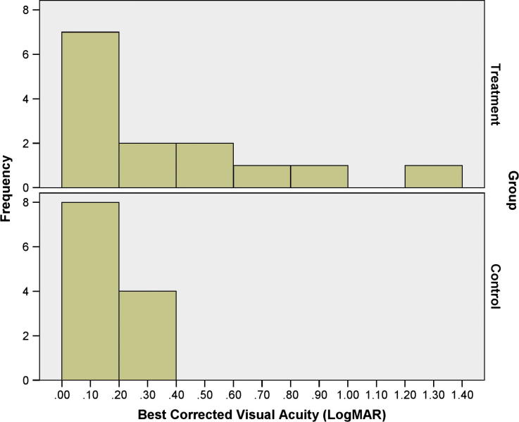
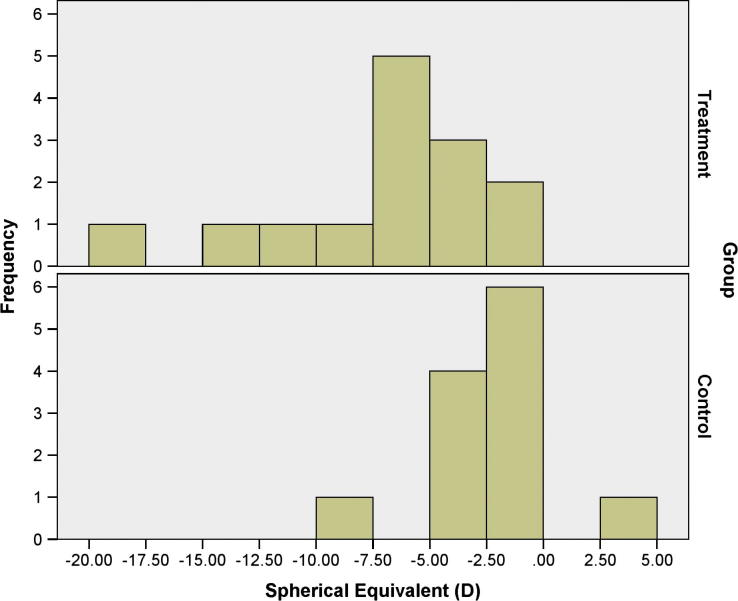
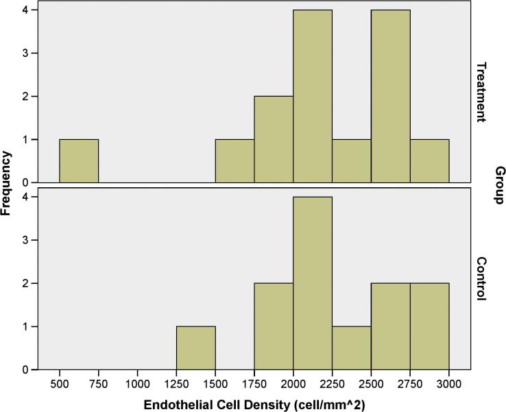
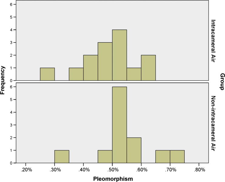
Results: The mean BCVA was 0.36 ± 0.36 logMAR in the treatment group and 0.17 ± 0.11 logMAR in the control group (p = 0.081), and the mean corneal thickness was 507.86 ± 62.69 μm in the treatment group and 525.67 ± 37.54 μm in the control group air (p = 0.399). Furthermore, the mean sphere was -5.14 ± 4.17D and -1.02 ± 3.29D, the mean cylinder was -3.16 ± 2.20D and -2.88 ± 1.21D, and the mean SEQ was -6.72 ± 4.66D and -2.46 ± 3.14D and in the treatment and control groups respectively (p = 0.011, 0.693, and 0.013). As to morphometric features, the mean ECD was 2176.76 ± 549.18 cell/mm(2) and 2257.30 ± 436.12 cell/mm(2) in the treatment and control groups respectively (p = 0.686), and the mean pleomorphism 0.48 ± 0.09 and 0.54 ± 0.10 in the treatment and control groups respectively (p = 0.139). In contrast, the mean polymegathism was 0.37 ± 0.06 and 0.31 ± 0.05 in the treatment and control groups respectively (p = 0.009).
Conclusion: The presence of air inside the anterior chamber for a short term may not cause further endothelial cell loss and can be safely performed to prevent postoperative Descemet Membrane detachment in case of micro perforations.
Keywords: Endothelial cell density; Keratoconus; Keratoplasty; Pleomorphism; Polymegathism.
Figures
Similar articles
-
Comparison of descemet-on versus descemet-off deep anterior lamellar keratoplasty.Cornea. 2013 Nov;32(11):1437-40. doi: 10.1097/ICO.0b013e3182a48028. Cornea. 2013. PMID: 24055906
-
Comparison of outcomes and complications of deep anterior lamellar keratoplasty and penetrating keratoplasty performed in a large group of patients with keratoconus.Int Ophthalmol. 2018 Jun;38(3):985-992. doi: 10.1007/s10792-017-0548-9. Epub 2017 May 22. Int Ophthalmol. 2018. PMID: 28534231
-
Prediction of Descemet Membrane Perforation During Deep Anterior Lamellar Keratoplasty in Patients With Keratoconus With Stromal Scar.Eye Contact Lens. 2018 Nov;44 Suppl 2:S176-S179. doi: 10.1097/ICL.0000000000000434. Eye Contact Lens. 2018. PMID: 29023312
-
Deep anterior lamellar keratoplasty techniques; predescemetic versus big bubble: Anterior segment optical coherence tomography study.J Fr Ophtalmol. 2020 Mar;43(3):222-227. doi: 10.1016/j.jfo.2019.08.004. Epub 2020 Jan 24. J Fr Ophtalmol. 2020. PMID: 31987676
-
Deep anterior lamellar keratoplasty in patients with keratoconus: big-bubble technique.Cornea. 2010 Feb;29(2):177-82. doi: 10.1097/ICO.0b013e3181af25b7. Cornea. 2010. PMID: 20023579
Cited by
-
Impact of Intraoperative Descemet Membrane Perforations on Deep Anterior Lamellar Keratoplasty Outcomes.J Ophthalmol. 2025 Jul 17;2025:4101770. doi: 10.1155/joph/4101770. eCollection 2025. J Ophthalmol. 2025. PMID: 40709101 Free PMC article. Review.
-
Corneal clarity measurements in patients with keratoconus undergoing either penetrating or deep anterior lamellar keratoplasty.Clin Ophthalmol. 2018 Mar 23;12:577-585. doi: 10.2147/OPTH.S157286. eCollection 2018. Clin Ophthalmol. 2018. PMID: 29615834 Free PMC article.
References
-
- Wang J., Zhao G., Xie L. Therapeutic effect of deep anterior lamellar keratoplasty for active or quiescent herpetic stromal keratitis. Graefes Arch Clin Exp Ophthalmol. 2012;250(8):1187–1194. - PubMed
-
- Fontana L., Parente G., Tassinari G. Clinical outcomes after deep anterior lamellar keratoplasty using the big-bubble technique in patients with keratoconus. Am J Ophthalmol. 2007;143(1):117–124. - PubMed
-
- Sarnicola V., Toro P., Sarnicola C. Long-term graft survival in deep anterior lamellar keratoplasty. Cornea. 2012;31(6):621–626. - PubMed
-
- Yanny Y.Y., Visser N., Schouten J.S. Endothelial cell loss and visual outcome of deep anterior lamellar keratoplasty versus penetrating keratoplasty: a randomized multicenter clinical trial. Ophthalmology. 2011;118(2):302–309. - PubMed
-
- Rodríguez-Calvo-de-Mora M., Quilendrino R., Ham L. Clinical outcome of 500 consecutive cases undergoing descemet’s membrane endothelial keratoplasty. Ophthalmology. 2015;122(3):464–470. - PubMed
LinkOut - more resources
Full Text Sources
Other Literature Sources
Research Materials
