. 2016 Jul-Aug;21(4):325-35.
doi: 10.1016/j.rpor.2014.09.002. Epub 2014 Oct 14.

Surgical management of skull base tumors

Affiliations

Surgical management of skull base tumors

Leonardo Rangel-Castilla et al. Rep Pract Oncol Radiother. 2016 Jul-Aug.

Abstract

Aim: To present a review of the contemporary surgical management of skull base tumors.

Background: Over the last two decades, the treatment of skull base tumors has evolved from observation, to partial resection combined with other therapy modalities, to gross total resection and no adjuvant treatment with good surgical results and excellent clinical outcomes.

Materials and methods: The literature review of current surgical strategies and management of skull base tumors was performed and complemented with the experience of Barrow Neurological Institute.

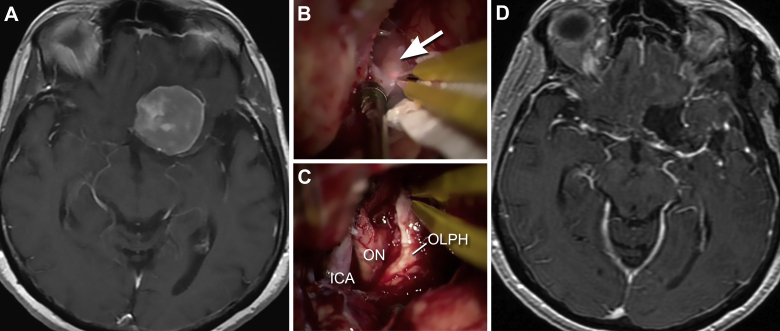
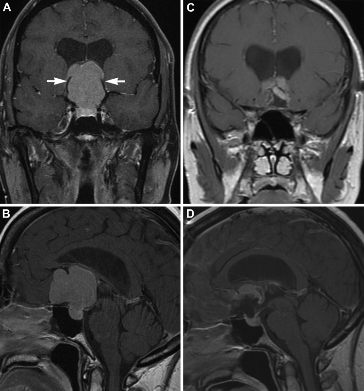
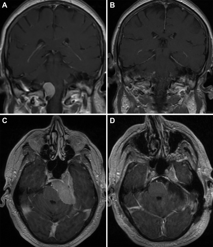
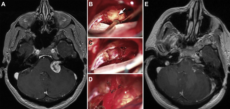
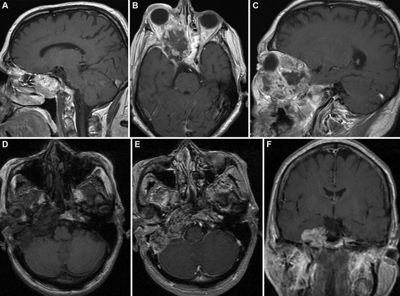
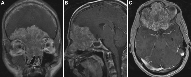
Results: Skull base tumors include meningiomas, pituitary tumors, sellar/parasellar tumors, vestibular and trigeminal schwannomas, esthesioneuroblastomas, chordomas, chondrosarcomas, and metastases. Surgical approaches include the modified orbitozygomatic, pterional, middle fossa, retrosigmoid, far lateral craniotomy, midline suboccipital craniotomy, and a combination of these approaches. The selection of an appropriate surgical approach depends on the characteristics of the patient and the tumor, as well as the experience of the neurosurgeon.

Conclusion: Modern microsurgical techniques, diagnostic imaging, intraoperative neuronavigation, and endoscopic technology have remarkably changed the concept of skull base surgery. These refinements have extended the boundaries of tumor resection with minimal morbidity.

Keywords: Meningoma; Neuroma; Schwannoma; Skull base; Skull base surgery.


Figures

Fig. 1
Fig. 1
Anterior skull base meningioma. A 65-year-old-female presented with left blurry vision. (A) T1-weighted axial magnetic resonance imaging (MRI) with gadolinium showing a left clinoidal meningioma. The patient underwent a left pterional craniotomy and tumor resection. (B and C) Intraoperative microscopic views of the tumor (arrow) pre- (B) and postresection (C). Even though the olfactory nerve was tightly adhered to the tumor, the nerve (OLPH) is preserved intact. Observe the anatomic relation of the olfactory nerve with the optic nerve (ON) and internal carotid artery (ICA). D, postoperative axial MRI demonstrates complete tumor resection. Used with permission from Barrow Neurological Institute.
Fig. 2
Fig. 2
Planum sphenoidale/tuberculum sellae meningioma. A 39-year-old-male presented with headaches, visual disturbances, and papilledema. A (coronal) and B (sagittal), T1-weighted MRIs with gadolinium show a large planum sphenoidale/tuberculum sellae meningioma with mass effect on both frontal lobes. The tumor is deforming the third ventricle, causing hydrocephalus. Also notice that both anterior cerebral arteries are stretched and deformed (A, arrows) by the tumor mass. The patient underwent a subfrontal craniotomy and interhemispheric approach for tumor resection. During the microsurgical resection, part of the tumor was extremely adhered to the roof of the third ventricle and fornices. This part of the tumor was not resected. C (coronal) and D (sagittal), postoperative MRIs show almost complete gross total resection. The patient will be managed conservatively with serial imaging. Used with permission from Barrow Neurological Institute.
Fig. 3
Fig. 3
Foramen magnum meningioma and petroclival meningioma. A and B, a 53-year-old-male presented with dizziness and vertigo. The T1-weighted coronal MRI (A) with gadolinium shows a right-side lateral foramen magnum meningioma causing mass effect on the brainstem and spinal cord. The patient underwent a right far lateral suboccipital craniotomy with C1 laminectomy and tumor resection. Postoperative coronal MRI (B) demonstrates complete surgical resection. C and D, a 63-year-old male with diplopia, ataxia, and decreased hearing. The T1-weighted axial MRI with gadolinium (C) demonstrates a large left petroclival meningioma causing brainstem compression. Notice that the tumor is infiltrating into the cavernous sinus. The patient underwent a left retrosigmoid craniotomy and tumor resection. The postoperative axial MRI (D) demonstrates gross total resection with excellent decompression of the brainstem. Used with permission from Barrow Neurological Institute.
Fig. 4
Fig. 4
Vestibular schwannoma. A 49-year-old-female presented with left side decreased hearing. (A) T1-weighted axial MRI with gadolinium shows a cystic cerebellopontine tumor causing mass effect on the cerebellum and brainstem. The patient underwent a left retrosigmoid craniotomy and tumor resection. (B–D) Intraoperative microscopic images show the tumor before resection (arrow) (B), after drilling the internal auditory canal (C), and after complete resection (D). Notice that the vestibular (V) and facial (F) cranial nerves were preserved. E, postoperative axial MRI demonstrates gross total resection. Used with permission from Barrow Neurological Institute.
Fig. 5
Fig. 5
Chordoma and chondrosarcoma. (A–C) A 67-year-old-male with a chordoma that was initially diagnosed at the age of 55 (A, sagittal MRI). He underwent multiple resections for tumor recurrences. Twelve years after the original diagnosis and 2 years after “complete” surgical resection, he presented with a large, infiltrative recurrence (B and C, axial and coronal MRIs, respectively). Notice the tumor extension into the ethmoid sinuses, cavernous sinus and right orbit. (D–F) 37-year-old-female with progressive right cranial nerve deficits (III, VI, VII and VIII) was found to have a skull base chondrosarcoma. Axial MRI pre- (D) and axial and coronal MRIs postcontrast (E and F) show the skull base mass lesion. These lesions required repeated extradural middle cranial fossa approaches for tumor control. Used with permission from Barrow Neurological Institute.
Fig. 6
Fig. 6
Esthesioneuroblastoma. A 25-year-old male presented with headaches, anosmia, and seizures. (A–C) Coronal, sagittal, and axial (respectively) T1-weighted MRIs with gadolinium demonstrate a large enhancing mass extending down into the ethmoidal sinuses and nasal cavity and extending up causing mass effect on both frontal lobes. The large mass occupies the entire anterior cranial base. Used with permission from Barrow Neurological Institute.

References

    1. Spetzler R.F., Sanai N. The quiet revolution: retractorless surgery for complex vascular and skull base lesions. J Neurosurg. 2012;116(2):291–300. - PubMed
    1. Waring A.J., Housworth C.M., Voorhies R.M., Douglas J.R., Walker C.F., Connolly S.E. A prototype retractor system designed to minimize ischemic brain retractor injury: initial observations. Surg Neurol. 1990;34(3):139–143. - PubMed
    1. Yamaguchi M., Jadhav V., Obenaus A., Colohan A., Zhang J.H. Matrix metalloproteinase inhibition attenuates brain edema in an in vivo model of surgically induced brain injury. Neurosurgery. 2007;61(5):1067–1075. discussion 1075–6. - PubMed
    1. Mackeith S.A., Kerr R.S., Milford C.A. Trends in acoustic neuroma management: a 20-year review of the oxford skull base clinic. J Neurol Surg B Skull Base. 2013;74(4):194–200. - PMC - PubMed
    1. Ammirati M.Z.H. Oncology. In: Brem H., editor. vol. 2. Elsevier Saunders; Philadelphia: 2011. (Youmans neurological surgery).

LinkOut - more resources