Introducing a Fresh Cadaver Model for Ultrasound-guided Central Venous Access Training in Undergraduate Medical Education
- PMID: 27330672
- PMCID: PMC4899071
- DOI: 10.5811/westjem.2016.3.30069
Introducing a Fresh Cadaver Model for Ultrasound-guided Central Venous Access Training in Undergraduate Medical Education
Abstract
Introduction: Over the past decade, medical students have witnessed a decline in the opportunities to perform technical skills during their clinical years. Ultrasound-guided central venous access (USG-CVA) is a critical procedure commonly performed by emergency medicine, anesthesia, and general surgery residents, often during their first month of residency. However, the acquisition of skills required to safely perform this procedure is often deficient upon graduation from medical school. To ameliorate this lack of technical proficiency, ultrasound simulation models have been introduced into undergraduate medical education to train venous access skills. Criticisms of simulation models are the innate lack of realistic tactile qualities, as well as the lack of anatomical variances when compared to living patients. The purpose of our investigation was to design and evaluate a life-like and reproducible training model for USG-CVA using a fresh cadaver.
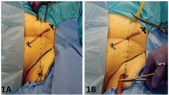
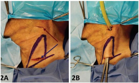
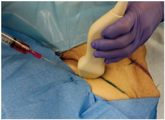
Methods: This was a cross-sectional study at an urban academic medical center. An 18-point procedural knowledge tool and an 18-point procedural skill evaluation tool were administered during a cadaver lab at the beginning and end of the surgical clerkship. During the fresh cadaver lab, procedure naïve third-year medical students were trained on how to perform ultrasound-guided central venous access of the femoral and internal jugular vessels. Preparation of the fresh cadaver model involved placement of a thin-walled latex tubing in the anatomic location of the femoral and internal jugular vein respectively.
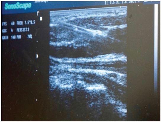
Results: Fifty-six third-year medical students participated in this study during their surgical clerkship. The fresh cadaver model provided high quality and lifelike ultrasound images despite numerous cannulation attempts. Technical skill scores improved from an average score of 3 to 12 (p<0.001) and procedural knowledge scores improved from an average score of 4 to 8 (p<0.001).
Conclusion: The use of this novel cadaver model prevented extravasation of fluid, maintained ultrasound-imaging quality, and proved to be an effective educational model allowing third-year medical students to improve and maintain their technical skills.
Figures
Similar articles
-
Ultrasound-guided procedures in medical education: a fresh look at cadavers.Intern Emerg Med. 2016 Apr;11(3):431-6. doi: 10.1007/s11739-015-1292-7. Epub 2015 Aug 15. Intern Emerg Med. 2016. PMID: 26276229
-
Assessment of ultrasound-guided procedures in preclinical years.Intern Emerg Med. 2017 Oct;12(7):1025-1031. doi: 10.1007/s11739-016-1525-4. Epub 2016 Aug 26. Intern Emerg Med. 2017. PMID: 27565985
-
Assessment of a new model for femoral ultrasound-guided central venous access procedural training: a pilot study.Acad Emerg Med. 2010 Jan;17(1):88-92. doi: 10.1111/j.1553-2712.2009.00626.x. Epub 2009 Dec 9. Acad Emerg Med. 2010. PMID: 20003122
-
The PASS Course: Practical Anatomy and Surgical Skills Course as a Pragmatic Primer for Senior Medical Students Pursuing Surgical Residency.Am Surg. 2023 May;89(5):1974-1979. doi: 10.1177/00031348211054551. Epub 2021 Nov 12. Am Surg. 2023. PMID: 34766513 Review.
-
[Ultrasound training in emergency and intensive care medicine : Integration already in medical school?].Med Klin Intensivmed Notfmed. 2019 Sep;114(6):519-524. doi: 10.1007/s00063-019-0550-2. Epub 2019 Mar 4. Med Klin Intensivmed Notfmed. 2019. PMID: 30830291 Review. German.
Cited by
-
Teaching medical students ultrasound-guided needle aspiration of synthetic cysts: effect of a formalin-embalmed cadaver simulation model.BMC Med Educ. 2024 Aug 27;24(1):931. doi: 10.1186/s12909-024-05907-z. BMC Med Educ. 2024. PMID: 39192224 Free PMC article. Clinical Trial.
-
Cadaver-based Necrotizing Fasciitis Model for Medical Training.Cureus. 2017 Apr 14;9(4):e1168. doi: 10.7759/cureus.1168. Cureus. 2017. PMID: 28507840 Free PMC article.
-
Comparison of In-person VS remote learning modalities for ultrasound-guided knee arthrocentesis training using formalin-embalmed cadavers.BMC Med Educ. 2024 Sep 7;24(1):974. doi: 10.1186/s12909-024-05930-0. BMC Med Educ. 2024. PMID: 39244572 Free PMC article. Clinical Trial.
-
Ultrasonography in undergraduate medical education: a comprehensive review and the education program implemented at Jichi Medical University.J Med Ultrason (2001). 2022 Apr;49(2):217-230. doi: 10.1007/s10396-021-01178-z. Epub 2022 Jan 16. J Med Ultrason (2001). 2022. PMID: 35034230 Free PMC article. Review.
-
Improving the quality of training paramedics by means of cadavers - a pilot study.BMC Med Educ. 2021 Jan 21;21(1):67. doi: 10.1186/s12909-021-02498-x. BMC Med Educ. 2021. PMID: 33494736 Free PMC article.
References
-
- Morton J, Anderson L, Frame F, et al. Back to the future: teaching medical students clinical procedures. Med Teach. 2006;28:723–8. - PubMed
-
- Talbott VA, Marks JA, Bodzin AS, et al. Technical skills acquisition in surgery-bound senior medical students: an evaluation of student assertiveness. J Surg Educ. 2012;69:529–35. - PubMed
-
- Fox RA, Ingham Clark CL, Scotland AD, et al. A study of pre-registration house officers’ clinical skills. Med Educ. 2000;34:1007–12. - PubMed
-
- Yudkowsky R, Loy G, York J. Ensuring medical student competency in basic procedural skills. Med Educ. 2005;39:515–6. - PubMed
-
- Evans DE, Wood DF, Roberts CM. The effect of an extended hospital induction on perceived confidence and assessed clinical skills of newly qualified pre-registration house officers. Med Educ. 2004;38:998–1001. - PubMed
MeSH terms
LinkOut - more resources
Full Text Sources
Other Literature Sources
Miscellaneous