. 2016 May 27;7(3):254-66.
doi: 10.14336/AD.2016.0118. eCollection 2016 May.

Failure of Elevating Calcium Induces Oxidative Stress Tolerance and Imparts Cisplatin Resistance in Ovarian Cancer Cells

Affiliations

Failure of Elevating Calcium Induces Oxidative Stress Tolerance and Imparts Cisplatin Resistance in Ovarian Cancer Cells

Liwei Ma et al. Aging Dis. .

Abstract

Cisplatin is a commonly used chemotherapeutic drug, used for the treatment of malignant ovarian cancer, but acquired resistance limits its application. There is therefore an overwhelming need to understand the mechanism of cisplatin resistance in ovarian cancer, that is, ovarian cancer cells are insensitive to cisplatin treatment. Here, we show that failure of elevating calcium and oxidative stress tolerance play key roles in cisplatin resistance in ovarian cancer cell lines. Cisplatin induces an increase in oxidative stress and alters intracellular Ca(2+) concentration, including cytosolic and mitochondrial Ca(2+) in cisplatin-sensitive SKOV3 cells, but not in cisplatin-resistant SKOV3/DDP cells. Cisplatin induces mitochondrial damage and triggers the mitochondrial apoptotic pathway in cisplatin-sensitive SKOV3 cells, but rarely in cisplatin-resistant SKOV3/DDP cells. Inhibition of calcium signaling attenuates cisplatin-induced oxidative stress and intracellular Ca(2+) overload in cisplatin-sensitive SKOV3 cells. Moreover, in vivo xenograft models of nude mouse, cisplatin significantly reduced the growth rates of tumors originating from SKOV3 cells, but not that of SKOV3/DDP cells. Collectively, our data indicate that failure of calcium up-regulation mediates cisplatin resistance by alleviating oxidative stress in ovarian cancer cells. Our results highlight potential therapeutic strategies to improve cisplatin resistance.

Keywords: calcium; cisplatin resistance; ovarian cancer; oxidative stress.


Figures

Figure 1.
Figure 1.. Cisplatin inhibits proliferation and induces cell death of ovarian cancer cells
(A) SKOV3 and SKOV3/DDP cells were treated with varying doses of cisplatin for 24 or 48 h. Cell viability was determined by the MTT assay. Data are presented as means ± SD, n=3. (B) Cells were treated with 6 μg/ml cisplatin for 0 h and 16 h, and then stained with Hoechst 33342 and PI. Then, cells were observed by confocal microscopy (bar, 20 μm). (C) Quantitation of cell death ratio. Data are presented as means ± SD, n=3, **P<0.01 vs. control.
Figure 2.
Figure 2.. Alteration of cytosolic Ca2+ is significantly induced in SKOV3 cells by cisplatin or ATP, but not in SKOV3/DDP cells
(A) Cells were treated with ATP and time-lapse imaging was used to detect changes in cytosolic Ca2+ levels. Data were obtained by confocal laser microscopy. (B) After cells treated with 6 μg/mL cisplatin for 0 h, 8 h and 16 h, cytosolic Ca2+ levels was detected by Fluo-4/AM (bar, 20 μm).
Figure 3.
Figure 3.. Cisplatin or ATP treatment induces mitochondrial Ca2+ influx in SKOV3, but not SKOV3/DDP cells
(A) Both cell lines were treated with ATP and time-lapse scanning was used to detect the changes of mitochondrial Ca2+. Data were obtained by confocal laser microscopy. (B) After cells treated with 6 μg/ml cisplatin for 0 h, 8 h and 16 h, mitochondrial Ca2+ was detected by confocal microscopy (bar, 20 μm).
Figure 4.
Figure 4.. Mitochondrial Ca2+ overload induces cell apoptosis through mitochondrial-dependent pathway in SKOV3 cells
(A) Representative transmission electron microscopy photomicrographs of both cell lines treated with 6μg/ml cisplatin for 8 h. Mitochondrial morphologies are normal in control cells (1,500x). Exposure to 6μg/ml cisplatin for 8 h resulted in mitochondrial damage (1,500x; arrows indicate mitochondrial damage). (B) Western blot analysis of cytosolic cytochrome c, caspase-3, and cleaved caspase-3 expression in cells treated with cisplatin for 0 h, 8 h, and 16 h. (C) Quantification of cytosolic cytochrome c and cleaved caspase-3 protein. Data are presented as means ± SD, n=3. **P<0.01 vs. control.
Figure 5.
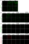
Figure 5.. Inhibition of cisplatin-induced cytosolic Ca2+ influx and mitochondrial Ca2+ overload reduces intracellular ROS production in SKOV3 cells
(A) Cells were treated with 6 μg/ml cisplatin for 0 h, 8 h and 16 h, and stained with DCFH-DA (bar, 20 μm). (B) After cells treated with 6 μg/ml cisplatin with or without 2-APB (50 μM) and BAPTA/AM (30 μM) for 16 h, cells were stained with DCFH-DA (bar, 20 μm). (C) After the same treatment with (A), cytosolic Ca2+ levels were detected by Fluo-4/AM and mitochondrial Ca2+ was detected by Rhod-2/AM (bar, 20 μm).
Figure 6.
Figure 6.. Inhibition of cisplatin-induced cytosolic Ca2+ influx and mitochondrial Ca2+ overload protect SKOV3 cells from cisplatin-induced apoptosis
(A) After cells treated with 6 μg/ml cisplatin with or without 2-APB (50 μM) and BAPTA/AM (30μM) for 16 h, cells were then stained with Annexin-V. Data are presented as the mean ± SD, n = 3. (B) After the same treatment with (A), the expression of caspase-3, cleaved caspase-3, and cytosolic cytochrome c in both cell lines is detected by western blotting. (C) Quantitation of cleaved caspase-3, and cytosolic cytochrome c protein levels. Data are presented as the mean ± SD, n = 3. **P < 0.01 vs. control, #P < 0.05 vs. cisplatin.
Figure 7.
Figure 7.. Cisplatin displays anti-tumor activity in xenograft mouse models bearing tumors originating from SKOV3 cells, but not SKOV3/DDP cells
(A) The average volume of tumor originating from SKOV3 cells were obviously diminished by cisplatin, but not in SKOV3/DDP cells. (B) BALB/c femal nude mice subcutaneous transplant tumor model was established using SKOV3 and SKOV3/DDP cells. After treatment with cisplatin, tumors in mice were excised and photographed for each group. Data are presented as the mean ± SD, n = 3. **P < 0.01 vs. control.

References

    1. Mei L, Chen H, Wei DM, Fang F, Liu GJ, Xie HY, Wang X, Zou J, Han X, Feng D (2013). Maintenance chemotherapy for ovarian cancer. Cochrane Database Syst Rev, 6:CD007414. - PMC - PubMed
    1. Pils D, Bachmayr-Heyda A, Auer K, Svoboda M, Auner V, Hager G, Obermayr E, Reiner A, Reinthaller A, Speiser P et al. (2014). Cyclin E1 (CCNE1) as independent positive prognostic factor in advanced stage serous ovarian cancer patients - a study of the OVCAD consortium. Eur J Cancer, 50(1):99-110. - PubMed
    1. Shank JJ, Yang K, Ghannam J, Cabrera L, Johnston CJ, Reynolds RK, Buckanovich RJ (2012). Metformin targets ovarian cancer stem cells in vitro and in vivo. Gynecol Oncol, 127(2):390-397. - PMC - PubMed
    1. Agarwal R, Kaye SB (2003). Ovarian cancer: strategies for overcoming resistance to chemotherapy. Nat Rev Cancer, 3(7):502-516. - PubMed
    1. Tummala MK, McGuire WP (2005). Recurrent ovarian cancer. Clin Adv Hematol Oncol, 3(9):723-736. - PubMed

LinkOut - more resources