

Single Operation to Repair Multifocal Cerebrospinal Fluid Fistulae Following Gunshot Wound: A Case Report
- PMID: 27330926
- PMCID: PMC4914715
- DOI: 10.1055/s-0036-1584281
Single Operation to Repair Multifocal Cerebrospinal Fluid Fistulae Following Gunshot Wound: A Case Report
Abstract
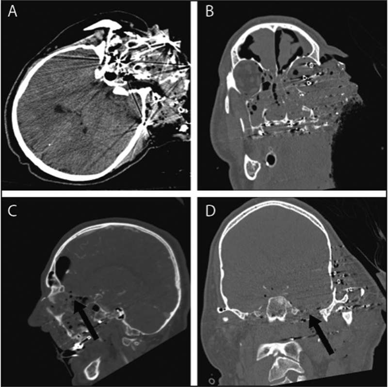
Introduction Traumatic cerebrospinal fluid (CSF) fistulae can be a challenging neurosurgical disease, often requiring complicated surgical intervention. Case Presentation A 54-year-old man presented with a gunshot wound to the head with complex injury to the skull base and significant CSF leakage from multiple sites. A single surgery was performed using a combined Neurosurgery, Neurotology, and Rhinology team, which was successful in repairing the multiple skull base defects and preventing further CSF leak. Discussion Trauma to the skull base is a common inciting factor for the development of CSF fistulae. Endoscopic approaches are often preferred for repairing these defects, but craniotomy remains a viable option that may be required in more complex cases. A combined approach has not been described previously, but was successful for this severe multifocal defect. Conclusion A multidisciplinary approach allowed for a combined intervention that addressed both the anterior and middle fossae fistulae simultaneously. This limited the potential infectious complications of continued CSF leak and allowed for early rehabilitation.
Keywords: CSF leak; cerebrospinal fluid fistulae; endoscopic endonasal approach; skull base repair.
Conflict of interest statement
Figures


References
-
- Plein C T, Langerman A J, Redleaf M I. Bilateral middle cranial fossa encephaloceles presenting as conductive hearing loss. Ear Nose Throat J. 2013;92(12):E14–E16. - PubMed
-
- Yilmazlar S, Arslan E, Kocaeli H. et al.Cerebrospinal fluid leakage complicating skull base fractures: analysis of 81 cases. Neurosurg Rev. 2006;29(1):64–71. - PubMed
-
- Dalgic A, Okay H O, Gezici A R, Daglioglu E, Akdag R, Ergungor M F. An effective and less invasive treatment of post-traumatic cerebrospinal fluid fistula: closed lumbar drainage system. Minim Invasive Neurosurg. 2008;51(3):154–157. - PubMed
-
- Liao W, Lin S H, Huang H T. Traumatic nasal cerebrospinal fluid fistula: report of 86 cases [in Chinese] J First Mil Med Univ. 2003;23(6):629–630. - PubMed
-
- Park J I, Strelzow V V, Friedman W H. Current management of cerebrospinal fluid rhinorrhea. Laryngoscope. 1983;93(10):1294–1300. - PubMed
Publication types
LinkOut - more resources
Full Text Sources
Other Literature Sources