. 2016 Jun 22;11(6):e0157641.
doi: 10.1371/journal.pone.0157641. eCollection 2016.

The Effect of Covalently-Attached ATRP-Synthesized Polymers on Membrane Stability and Cytoprotection in Human Erythrocytes

Affiliations

The Effect of Covalently-Attached ATRP-Synthesized Polymers on Membrane Stability and Cytoprotection in Human Erythrocytes

William P Clafshenkel et al. PLoS One. .

Abstract

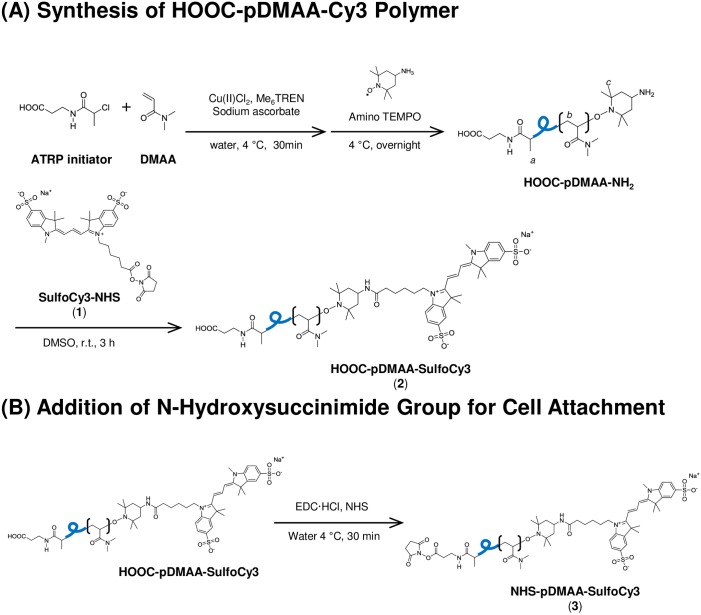
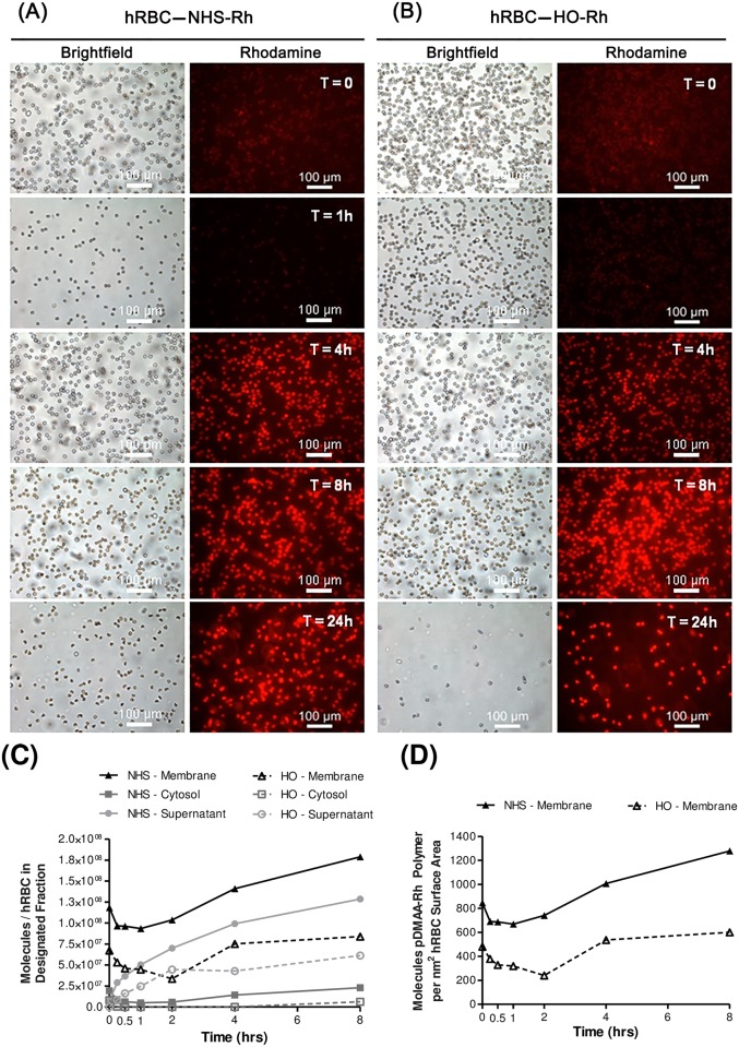
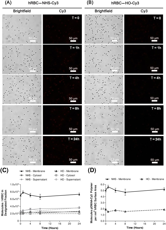
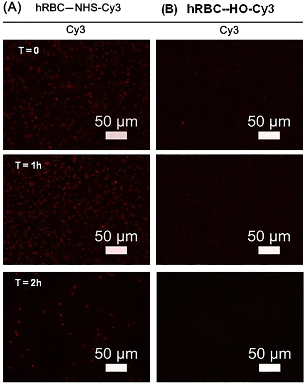
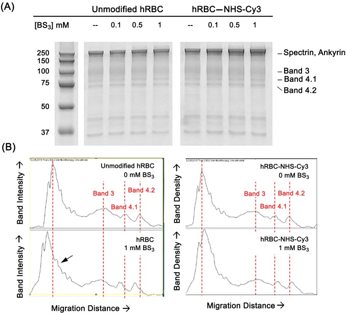
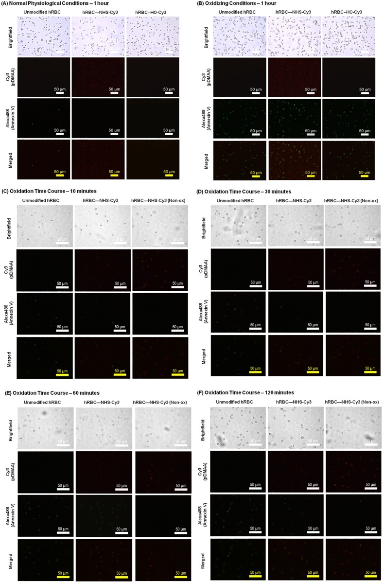
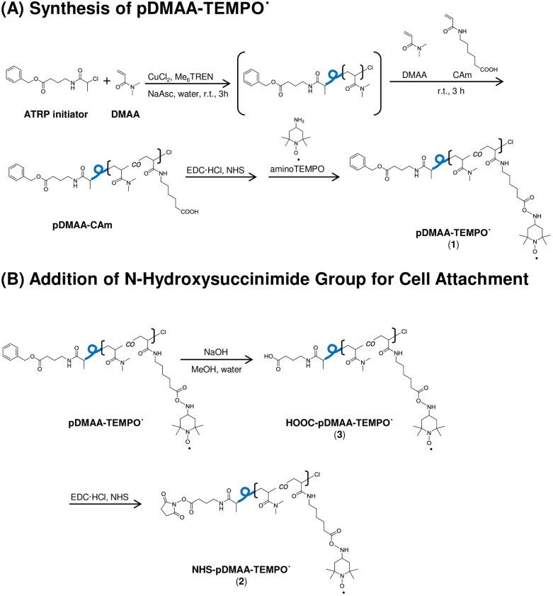
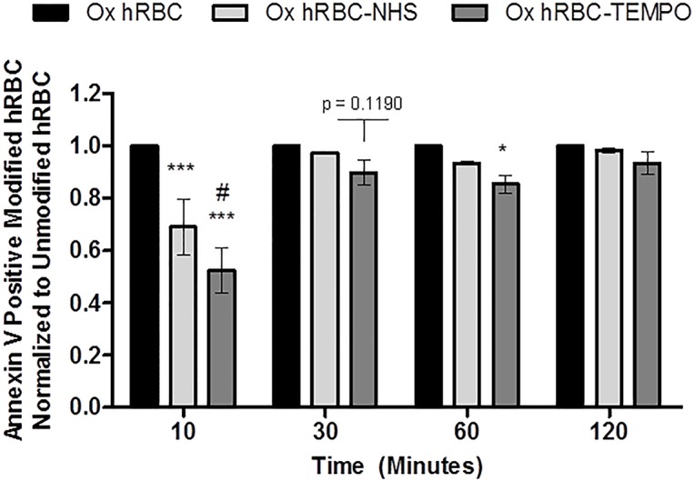
Erythrocytes have been described as advantageous drug delivery vehicles. In order to ensure an adequate circulation half-life, erythrocytes may benefit from protective enhancements that maintain membrane integrity and neutralize oxidative damage of membrane proteins that otherwise facilitate their premature clearance from circulation. Surface modification of erythrocytes using rationally designed polymers, synthesized via atom-transfer radical polymerization (ATRP), may further expand the field of membrane-engineered red blood cells. This study describes the fate of ATRP-synthesized polymers that were covalently attached to human erythrocytes as well as the effect of membrane engineering on cell stability under physiological and oxidative conditions in vitro. The biocompatible, membrane-reactive polymers were homogenously retained on the periphery of modified erythrocytes for at least 24 hours. Membrane engineering stabilized the erythrocyte membrane and effectively neutralized oxidative species, even in the absence of free-radical scavenger-containing polymers. The targeted functionalization of Band 3 protein by NHS-pDMAA-Cy3 polymers stabilized its monomeric form preventing aggregation in the presence of the crosslinking reagent, bis(sulfosuccinimidyl)suberate (BS3). A free radical scavenging polymer, NHS-pDMAA-TEMPO˙, provided additional protection of surface modified erythrocytes in an in vitro model of oxidative stress. Preserving or augmenting cytoprotective mechanisms that extend circulation half-life is an important consideration for the use of red blood cells for drug delivery in various pathologies, as they are likely to encounter areas of imbalanced oxidative stress as they circuit the vascular system.


Conflict of interest statement

Competing Interests: The authors have declared that no competing interests exist.

Figures

Fig 1
Fig 1. Schematic for the ATRP synthesis of a disulfoCy3-containing poly(DMAA) polymer.
Disulfo-Cy3 was added to polymer chains for fluorescence imaging of polymer-exposed cells (1). NHS functional group attachment facilitates cell attachment via covalent amide bond formation with surface proteins (3). Polymer chains lacking NHS functionality were used as a control for cell surface binding (2). The average molecular weight of polymer chains was approximately 7 kDa. Additional information regarding the synthesis of the polymers described in this study can be found in S1 File.
Fig 2
Fig 2. Human erythrocyte membrane engineering with NHS-pDMAA-Rh polymers.
NHS-pDMAA-Rh polymers exhibit significant non-specific binding and initial high-density fluorescence quenching. Human RBCs were modified with 182 μM NHS-pDMAA-Rh or 98 μM HO-pDMAA-Rh for 30 minutes at 37°C. (A) Representative images of NHS-pDMAA-Rh-exposed hRBC for each designated time point. (B) Representative images of HO-pDMAA-Rh-exposed hRBC for each designated time point. Epifluorescent images were capture after washing on a Leica inverted microscope at 20X. Scale bars measure 100 μm. (C) Supernatant, cytosolic, and membrane fractions were collected at 0, 1, 4, and 8 hours. Polymer retention and internalization was assessed by monitoring the relative fluorescence of each fraction over time and calculated as the number of polymer molecules per hRBC using a standard curve. (D) The number of polymer molecules per nm2 hRBC surface area was calculated using an average hRBC surface area of 140 μm2. Images were background corrected and the brightness/contrast for each channel was balanced using Image J software. n = 3.
Fig 3
Fig 3. Human erythrocyte membrane engineering with NHS-pDMAA-Cy3 polymers.
NHS-pDMAA-Cy3 polymers are retained on hRBC membranes over a 24 hour period compared to HO-pDMAA-Cy3 polymers, indicating cell surface reactivity. Human RBCs were modified with 100 μM NHS-pDMAA-Cy3 or HO-pDMAA-Cy3 for 30 minutes at 37°C. (A) Representative images of NHS-pDMAA-Cy3-exposed hRBC for each designated time point. (B) Representative images of HO-pDMAA-Cy3-exposed hRBC for each designated time point. Epifluorescent images were capture after washing on a Leica inverted microscope at 40X. Scale bars measure 50 μm. (C) Supernatant, cytosolic, and membrane fractions were collected at 0, 1, 4, 8, and 24 hours. Polymer retention and internalization was assessed by monitoring the relative fluorescence of each fraction over time and calculated as the number of polymer molecules per hRBC using a standard curve. (D) The number of polymer molecules per nm2 hRBC surface area was calculated using an average hRBC surface area of 140 μm2. Images were background corrected and the brightness/contrast for each channel was balanced using Image J software. n = 3.
Fig 4
Fig 4. Human ghost erythrocyte membrane engineering with NHS-pDMAA-Cy3 polymers.
Human erythrocyte ghosts can be surface modified with NHS-pDMAA-Cy3 and exhibit short-term retention of polymer. Ghost membranes were produced from intact hRBCs using hypotonic methods. Human RBC ghosts were exposed to either 100 μM NHS- or HO-pDMAA-Cy3 polymers for 30 minutes at 37°C. Epifluorescent images were captured after washing using an inverted Leica microscope at 40X. hRBC exposed to control polymer demonstrate negligible fluorescent signal (B) while those exposed to NHS-pDMAA-Cy3 exhibit strong fluorescence that diminishes over 2 hours (A). Representative images are shown for each time point. Scale bars measure 50 μm. Images were background corrected and the brightness/contrast for each channel was balanced using Image J software.
Fig 5
Fig 5. The effect of human erythrocyte membrane engineering with NHS-pDMAA-Cy3 polymers on Band 3 aggregation.
Human erythrocyte Band 3 protein is a major site of NHS-pDMAA-Cy3 modification supported by the absence of Band 3 aggregate formation in the presence of the protein cross-linker bis(sulfosuccinimidyl)suberate (BS3). (A) Isolated hRBC membranes for each group were run on 12% TGX polyacrylamide gels and stained with Imperial protein stain. Gel image truncated after 37 kDa molecular weight marker to highlight larger molecular weight membrane proteins. (B) Histogram plots of band intensity versus migration distance were plotted for each experimental group and concentration (0 mM and 1 mM BS3 concentrations shown). Dotted lines are used to represent peak intensity of specific protein bands in non-crosslinked hRBC membranes. Specifically, at the highest concentration of BS3 (1 mM) there is a noticeable decrease in monomeric Band 3 protein band intensity as well as a modest increase in higher molecular weight proteins gathering at the top of the gel (e.g., in the region of spectrin and ankyrin proteins) for unmodified hRBCs versus those modified with NHS-pDMAA-Cy3 polymers. The arrow represents Band 3 aggregates.
Fig 6
Fig 6. Effect of human erythrocyte membrane engineering with NHS-pDMAA-Cy3 polymers on membrane stability.
(A) Modification of hRBC with NHS-pDMAA-Cy3 polymers does not induce membrane destabilization as evidenced by the absence of PS-Annexin V signal under physiological conditions. Both unmodified and modified (100 μM polymer solution) hRBC were incubated in 1X PBS for one hour at 37°C. Annexin V-Alexa488 binding to externalized phosphatidylserine was used to monitor membrane destabilization and oxidative damage (green fluorescence). Cy3-modification demonstrated by red fluorescence. Epifluorescent images of PS-Annexin V and Cy3 channels captured under oil at 60x using an inverted Leica microscope. Individual channels were merged using Image J. Scale bars measure 50 μm. Images were background corrected and the brightness/contrast for each channel was balanced using Image J software. (B) Effect of human erythrocyte membrane engineering with NHS-pDMAA-Cy3 polymers on membrane damage during oxidant exposure. Modification of hRBC with NHS-pDMAA-Cy3 polymers may protect against membrane damage under oxidizing conditions demonstrated by a potential mitigation of phosphatidylserine-Annexin V signal. Both unmodified and modified (100 μM polymer solution) hRBC were incubated in 0.2 mM CuSO4/2.5 mM ascorbate solution for one hour at 37°C. Annexin V-Alexa488 binding to externalized phosphatidylserine was used to monitor membrane destabilization and oxidative damage (green fluorescence). Cy3-modification demonstrated by red fluorescence. Epifluorescent images of PS-Annexin V and Cy3 channels captured under oil at 60X using an inverted Leica microscope. Individual channels were merged using Image J. Scale bars measure 50 μm. Images were background corrected and the brightness/contrast for each channel was balanced using Image J software. (C-F). Time point analysis of the effect of human erythrocyte membrane engineering with NHS-pDMAA-Cy3 polymers on membrane damage during oxidant exposure. Modification of hRBC with NHS-pDMAA-Cy3 polymers may protect against membrane damage under oxidizing conditions demonstrated by a potential mitigation of phosphatidylserine-Annexin V signal over time. To further investigate the temporal regulation of these events, both unmodified and modified (100 μM polymer solution) hRBC were incubated in 0.2 mM CuSO4/2.5 mM ascorbate solution for 10 (C), 30 (D), 60 (E), or 120 (F) minutes at 37°C. Annexin V-Alexa488 binding to externalized phosphatidylserine was used to monitor membrane destabilization and oxidative damage (green fluorescence). Cy3-modification demonstrated by red fluorescence. Epifluorescent images of PS-Annexin V and Cy3 channels captured under oil at 60X using an inverted Leica microscope. Individual channels were merged using Image J. Scale bars measure 50 μm. Images were background corrected and the brightness/contrast for each channel was balanced using Image J software.
Fig 7
Fig 7. Schematic for the ATRP synthesis of a TEMPO-terminated poly(DMAA) polymer.
The stable free-radical nitroxide, 4-amino-2,2,6,6-tetramethylpiperidine-1-oxyl (TEMPO˙) was added to polymer chains to investigate its effects as a cytoprotective antioxidant attached to human red blood cells (1). NHS functional group attachment facilitates cell attachment via covalent amide bond formation with surface proteins (2). Polymer chains lacking NHS functionality were used as a control for cell surface binding (3). The average molecular weight of polymer chains was approximately 6.2kDa.
Fig 8
Fig 8. Effect of human erythrocyte membrane engineering with NHS-pDMAA-TEMPO˙ polymers on membrane damage during oxidant exposure over time.
Human erythrocytes modified with either NHS-pDMAA-Cy3 or NHS-pDMAA-TEMPO˙ demonstrated significant or nearly significant cytoprotective trends over time when exposed to oxidizing conditions (0.1 mM CuSO4/1.25 mM ascorbate solution). Results are normalized to unmodified, oxidized hRBCs for each time point. Oxidation in this assay is indirectly measured by monitoring externalized phosphatidylserine tagging by Annexin V-Alexa488 using flow cytometry. Cell populations were gated as either positive or negative for Annexin V-Alexa488 fluorescence. *** indicates significance, p < 0.001 compared to unmodified hRBCs; * indicates significance, p < 0.05 compared to unmodified hRBCs; # indicates significance, p < 0.05 compared to NHS-pDMAA-Cy3 modified hRBCs. NHS-pDMAA-TEMPO˙-modified hRBCs are nearly significantly different to unmodified hRBCs at 30 minutes (p = 0.1190). Results were analyzed by a two-way ANOVA with Fisher’s LSD post hoc test. Bars represent group means ± SEM. n = 3.

Similar articles

Cited by

References

    1. Muzykantov VR. Drug delivery by red blood cells: vascular carriers designed by mother nature. Expert opinion on drug delivery. 2010;7(4):403–27. 10.1517/17425241003610633 - DOI - PMC - PubMed
    1. Siems WG, Sommerburg O, Grune T. Erythrocyte free radical and energy metabolism. Clinical nephrology. 2000;53(1 Suppl):S9–17. - PubMed
    1. Szweda-Lewandowska Z, Krokosz A, Gonciarz M, Zajeczkowska W, Puchala M. Damage to human erythrocytes by radiation-generated HO* radicals: molecular changes in erythrocyte membranes. Free radical research. 2003;37(10):1137–43. - PubMed
    1. Lutz HU, Bogdanova A. Mechanisms tagging senescent red blood cells for clearance in healthy humans. Frontiers in physiology. 2013;4:387 10.3389/fphys.2013.00387 - DOI - PMC - PubMed
    1. Vissers MC, Carr AC, Chapman AL. Comparison of human red cell lysis by hypochlorous and hypobromous acids: insights into the mechanism of lysis. The Biochemical journal. 1998;330 (Pt 1):131–8. - PMC - PubMed

LinkOut - more resources