. 2016 Jul 19;7(29):46401-46418.
doi: 10.18632/oncotarget.10136.

RRD-251 enhances all-trans retinoic acid (RA)-induced differentiation of HL-60 myeloblastic leukemia cells

Affiliations

RRD-251 enhances all-trans retinoic acid (RA)-induced differentiation of HL-60 myeloblastic leukemia cells

Aaron S Wallace et al. Oncotarget. .

Abstract

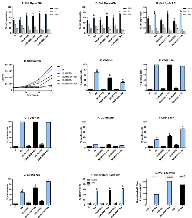
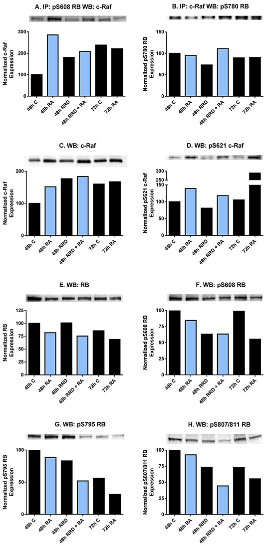
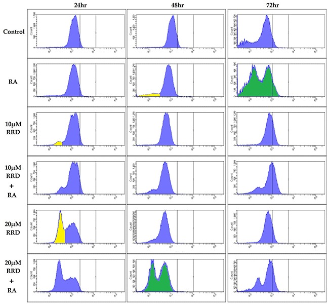
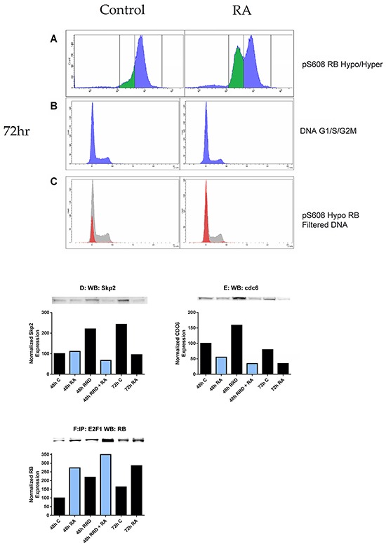
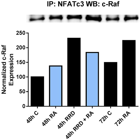
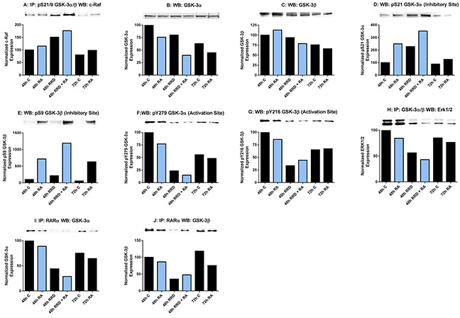
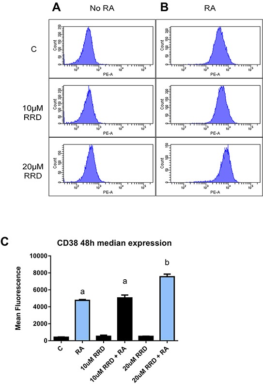
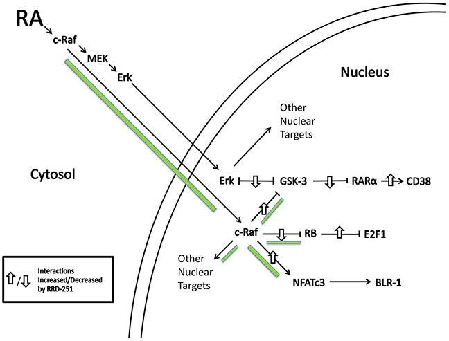
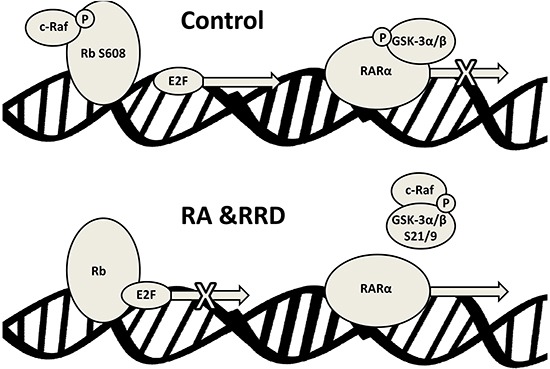
All-trans-retinoic acid (RA) is known to induce terminal granulocytic differentiation and cell cycle arrest of HL-60 cells. Responding to an RA-induced cytosolic signaling machine, c-Raf translocates to the nucleus, providing propulsion for RA-induced differentiation. This novel mechanism is not understood, but presumably reflects c-Raf binding with nuclear gene regulatory proteins. RRD-251 is a small molecule that prevents the interaction of c-Raf and RB, the retinoblastoma tumor suppressor protein. The involvement of c-Raf and RB in RA-induced differentiation motivates interest in the effects of combined RA and RRD-251 treatment on leukemic cell differentiation. We demonstrate that RRD-251 enhances RA-induced differentiation. Mechanistically, we find that nuclear translocated c-Raf associates with pS608 RB. RA causes loss of pS608 RB, where cells with hypophosphorylated S608 RB are G0/G1 restricted. Corroborating the pS608 RB hypophosphorylation, RB sequestration of E2F increased with concomitant loss of cdc6 expression, which is known to be driven by E2F. Hypophosphorylation of S608 RB releases c-Raf from RB sequestration to bind other nuclear targets. Release of c-Raf from RB sequestration results in enhanced association with GSK-3 which is phosphorylated at its S21/9 inhibitory sites. c-Raf binding to GSK-3 is associated with dissociation of GSK-3 and RARα, thereby relieving RARα of GSK-3 inhibition. RRD-251 amplifies each of these RA-induced events. Consistent with the posited enhancement of RARα transcriptional activity by RRD-251, RRD-251 increases the RARE-driven CD38 expression per cell. The RA/c-Raf/GSK-3/RARα axis emerges as a novel differentiation regulatory mechanism susceptible to RRD-251, suggesting enhancing RA-effects with RRD-251 in therapy.

Keywords: GSK-3; RRD-251; c-Raf; retinoblastoma protein (RB); all-trans retinoic acid (RA).


Conflict of interest statement

None of the authors have competing interests.

Figures

Figure 1
Figure 1. RRD-251 enhances RA-induced differentiation
HL-60 cells were untreated (C) or treated with 1 μM RA, 10 or 20 μM RRD-251, or RA plus 10 or 20 μM RRD-251 for the designated time points. The statistically significant changes are labeled a, b, c on the graphs. A. Cell cycle distribution (measured by flow cytometry with propidium iodide staining) at 24 hours after treatment indicates a dose-dependent enrichment of G0/G1 by RRD-251(C vs. 10 μM RRD-251 p<0.05; C vs. 20 μM RRD-251 p<0.001). B. At 48 hours post treatment, RA induces cell cycle arrest (p<0.0002) and cells treated with both 20 μM RRD-251 and RA exhibited even greater levels of cell cycle arrest than RA alone (p<0.005). C. By 72 hours after treatment, enrichment in G0/G1 is solely dependent on RA treatment (p<0.01). D. Cell density was measured for both treated and untreated cells at 0, 24, 48, and 72 hours. RRD-251 alone significantly inhibited growth in a dose dependent manner and enhances RA-induced growth inhibition. E. CD38 expression (assessed by flow cytometry with a PE-conjugated antibody) at 8 hours post treatment is induced by RA, but inhibited in dose dependent manner by RRD-251 (RA vs 10 μM RRD-251 + RA p=0.001; 10 μM RRD-251 + RA vs 20 μM RRD-251 p<0.05). F. At 24 hours post treatment, CD38 expression by RA is nearly 100% and is not inhibited by RRD-251 treatment. G. At 48 hours post treatment, CD38 expression by RA is nearly 100% and is not inhibited by RRD-251 treatment. H. CD11b expression (assessed by flow cytometry with an APC-conjugated antibody) at 24 hours post treatment is slightly induced by RA with no affect with RRD-251 treatment. I. At 48 hours, RA-induced expression of CD11b increases and is augmented with RRD-251 treatment (RA vs 10 μM RRD-251 + RA p<<0.05; 10 μM RRD-251 + RA vs 20 μM RRD-251 p<0.0005). J. At 72 hours, RRD-251 again enhances RA-induced expression of CD11b (RA vs 20 μM RRD-251 + RA p<0.05). K. Respiratory burst, measured by flow cytometry of DCF stained cells, is significantly enhanced at 72 hours by co-treatment compared to RA alone (p=0.021). L. Expression of p47phox was measured by Western blotting at 48 hours for control, RA, 20 μM RRD-251, and RA+ 20 μM RRD-251 and at 72 hours for control and RA. RRD-251 significantly augments the RA-induced expression, where there is greater expression in the RRD-251 plus RA samples at 48 hours than the RA samples at 72 hours. All the experiments were performed three times (three biological repeats).
Figure 2
Figure 2. RA and RRD-251 promote transient changes in the c-Raf-RB interaction and their specific phosphorylation
300 μg of pre-cleared nuclear lysate collected 48 or 72 hours post treatment were incubated overnight with 1 μg of the precipitating antibody with magnetic beads and resolved on 7.5% polyacrylamide gels. A. A pS608 RB antibody was used as the precipitating antibody and the blot was probed by overnight incubation with c-Raf primary antibody. RA promotes a transient nuclear interaction between c-Raf and pS608 RB at 48, but not at 72 hours post treatment. The presence of RRD-251 disrupts the RA-promoted interaction at 48 hours, where co-treated samples at 48hours post treatment resemble 72 hours 1 μM RA-treated samples. B. A c-Raf antibody was used as the precipitating antibody and the blot was probed with pS780 RB primary antibody. There is not any significant modulation in the interaction between c-Raf and pS780 RB with either drug treatment. C. Changes in the specific phosphorylation of c-Raf and RB were assessed by Western blotting. Nuclear lysates collected 48 or 72 hours after treatment were resolved on 7.5% polyacrylamide gels. RRD-251 samples were only collected 48 hours after treatment. 25 μg of protein was loaded per well. RA induces the nuclear translocation of c-Raf, which is augmented by RRD-251 treatment. D. RA promotes the phosphorylation of c-Raf at serine 621 at 48 and 72 hours after treatment, which is not enhanced in co-treated samples. E. RA promotes the downregulation of total RB. F. RA promotes a decrease in pS608 RB levels at 48 and 72 hours, but most significantly at 72 hours. This decrease is amplified with RRD-251 treatment. G-H. Westerns of pS795 RB and pS807/811 RB indicate that RA induces the global dephosphorylation of RB at 72 hours, which is observed in co-treated samples at 48 hours post treatment.
Figure 3
Figure 3. RB serine 608 phosphorylation is modulated by RA and RRD-251
Cells were collected at 24, 48, and 72 hours after experiment initiation and fixed for 7 minutes in 100 μL 2% para-formaldehyde PBS solution and permeabilized with 900 μL ice-cold methanol. Following several washings with PBS, samples were incubated with pS608 RB primary antibody overnight. The samples were incubated with a FITC goat anti-rabbit secondary antibody and analyzed by flow cytometry. Cells were either control (Row 1), 1 μM RA (Row 2), 10 μMRRD-251 (Row 3), 10 μMRRD-251+1 μM RA (Row 4), 20 μMRRD-251 (Row 5), or 20 μMRRD-251+ 1 μM RA (Row 6) and collected at 24, 48, and 72 hours. At 24 hours, RRD-251 induces a population with hypophosphorylated serine 608 RB. At 48 hours, the population is present in RA and RRD-251 samples. RA induces the population at 72 hours. Comparing the two green histograms shows that RA induces a hypo and hyper pS608 RB by 72 hours whereas RA+RRD-251 does it by 48 hours. The yellow marks hypo phosphorylated pS608 RB. Three biological repeats were analyzed.
Figure 4
Figure 4. RB serine 608 phosphorylation corresponds to G0/G1
Cells were fixed and permeabilized in the same method as before, but incubated in hypotonic PI solution following the removal of the methanol/para-formaldehyde solution. Compensation of 2.0% was used to correct for spillover of PI fluorescence into the FITC channel. A. 1 μM RA induces the formation of a population with hypophosphorylated serine 608 RB at 72 hours. B. The DNA histogram (cell cycle distribution) of control and RA cells at 72 hours is depicted. C. The hypophosphorylated population is overwhelmingly in G0/G1 (red histogram) as compared to the hyperphosphorylated serine 608 RB population (grey histogram). D-E. Changes in expression of Skp2 and cdc6 were measured by Western blotting. Nuclear lysates collected 48 or 72 hours after treatment were resolved on 7.5% polyacrylamide gels. 25 μg of protein was loaded per well. D. RA promotes a decrease in nuclear Skp2 at 72 hours, which is observed in co-treated samples at 48 hours. E. RA promotes a decrease in nuclear cdc6 at both 48 and 72 hours. Levels of cdc6 are lowest in co-treated samples at 48 hours. F. The RB-E2F1 interaction was assessed by immunoprecipitation. 300 μg of pre-cleared nuclear lysate collected 48 or 72 hours post treatment were incubated overnight with 1 μg of the precipitating antibody with magnetic beads and resolved on 7.5% polyacrylamide gels. RA promotes the interaction between RB and E2F1 at both 48 and 72 hours after treatment as compared to control. RRD-251 alone induces this interaction and there is an additive effect with co-treatment of RA at 48 hours. Co-treated samples at 48 hours exhibit higher levels of the interaction than 72 hours RA-treated cells.
Figure 5
Figure 5. RRD-251 increases the RA-induced c-Raf-NFATc3 interaction
The c-Raf-NFATc3 interaction was assessed by immunoprecipitation. 300 μg of pre-cleared nuclear lysate collected 48 or 72 hours post treatment were incubated overnight with 1 μg of the precipitating antibody with magnetic beads and resolved on 7.5% polyacrylamide gels. RA induces the c-Raf-NFATc3 interaction at 48 and 72 hours post treatment, with greater binding at 72 hours. Adding RRD-251 causes greater binding at 48 hours compared to RA-alone.
Figure 6
Figure 6. RRD-251 augments RA regulation of GSK-3 activity and interactions
GSK-3 phosphorylation was assessed by western blotting and its interactions with c-Raf and RARα were assessed by immunoprecipitation. Nuclear lysates collected 48 or 72 hours after treatment were resolved on 7.5% polyacrylamide gels. RRD-251 samples were only collected 48 hours after treatment. 25 μg of protein was loaded per well for western blotting. For immunoprecipitation, 300 μg of pre-cleared nuclear lysate collected 48 or 72 hours post treatment were incubated overnight with 1 μg of the precipitating antibody with magnetic beads and resolved on 7.5% polyacrylamide gels. A. Immunoprecipitation of pS21/9 GSK-3α/β probed for c-Raf. RA induces a novel interaction between c-Raf and GSK-3 apparent at 48 and 72 hours. B. Nuclear GSK-3α levels were assessed by western blotting. Decrease in expression induced by RA is augmented by RRD-251 co-treatment. C. Nuclear GSK-3β levels were assessed by western blotting. Expression in RA-treated cells is decreased by adding RRD-251, as was for GSK-3α. D-G. Western blots of GSK-3α and β phosphorylated at inhibitory pS21/9 and activating pY279/216 sites. At 48 and 72 hours post treatment, RA induces the inhibitory phosphorylation of serine 21 and serine 9 of GSK-3. RA also slightly inhibits the activating phosphorylation of GSK-3 at tyr279 and tyr216. Addition of RRD-251 enhanced these effects. H. Immunoprecipitation of GSK-3α/β probed for ERK1/2. RRD-251 further reduces the diminished interaction between GSK-3 and ERK1/2 in RA-treated cells. I-J. Immunoprecipitation of RARα probed for GSK-3α and β RRD-251 further diminishes the GSK-3-RARα interaction in RA-treated cells, which is known to inhibit RARα transcriptional activity. The addition of RRD-251 significantly further reduces this interaction. GSK-3 is known to bind and inhibit RARα. Three biological repeats were performed and the trend for changes in expression levels are consistent among the repeats.
Figure 7
Figure 7. RRD-251 enhances median CD38 expression
Median expression of CD38 per cell was assessed by flow cytometry using a PE-conjugated antibody) at 48 hours in control, RA-treated and RA plus RRD-251- treated cells. A. Histograms of CD38 expression in cells without RA show negative peak. B. Histograms of CD38 expression in samples treated with RA show a dose dependent increase in median expression per cell with the addition of RRD-251. (The histograms top, middle, bottom are 1 μM RA, 10 μM RRD-251+1 μM RA, 20 μM RRD-251+1 μM RA). C. Bar graph of median CD38 expression per cell showing that addition of RRD-251 to RA treated cells significantly increases expression (p=0.0041). Groups with different letters (a or b) are significantly different; the cells treated with 20 μMRRD-251+ 1 μM RA have significantly more CD38 than cells treated with just RA.
Figure 8
Figure 8. RA induces c-Raf nuclear translocation and binding to nuclear proteins
RRD-251 augments those effects. Green accent shows flow of c-Raf. Up/down arrows show effects shown to be affected by adding RRD-251.
Figure 9
Figure 9. Schematic representation of the nuclear events induced by RA and RRD-251 co-treatment on RAREs

Similar articles

Cited by

References

    1. Cull EH, Altman JK. Contemporary treatment of APL. Curr Hematol Malig Rep. 2014;9:193–201. - PMC - PubMed
    1. Brown G, Hughes P. Retinoid differentiation therapy for common types of acute myeloid leukemia. Leuk Res Treatment. 2012;2012:939021. - PMC - PubMed
    1. Bunaciu RP, Yen A. Retinoid Chemoprevention: Who Can Benefit? Curr Pharmacol Rep. 2015;1:391–400. - PMC - PubMed
    1. Tallman MS, Gilliland DG, Rowe JM. Drug therapy for acute myeloid leukemia. Blood. 2005;106:1154–1163. - PubMed
    1. Kohrt HE, Coutre SE. Optimizing therapy for acute myeloid leukemia. J Natl Compr Canc Netw. 2008;6:1003–1016. - PubMed

MeSH terms