

Co-expression of wild-type FLT3 attenuates the inhibitory effect of FLT3 inhibitor on FLT3 mutated leukemia cells
- PMID: 27331411
- PMCID: PMC5216920
- DOI: 10.18632/oncotarget.10147
Co-expression of wild-type FLT3 attenuates the inhibitory effect of FLT3 inhibitor on FLT3 mutated leukemia cells
Abstract
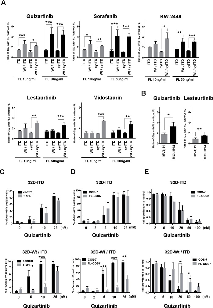
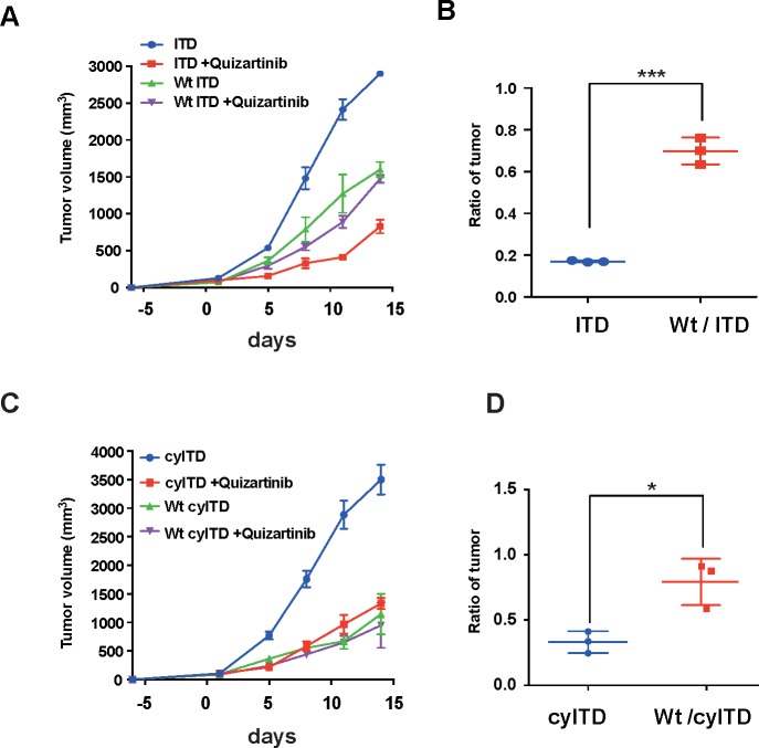
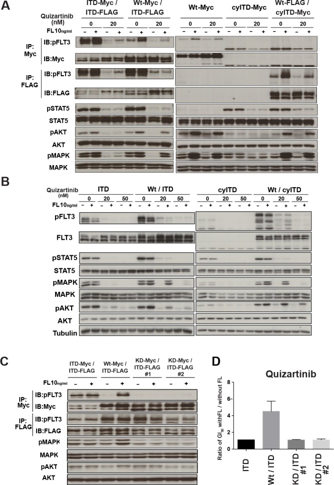
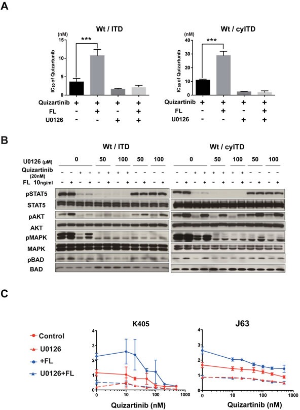
FLT3 mutation is found in about 30% of acute myeloid leukemia (AML) patients and is associated with a poor prognosis. Several FLT3 inhibitors are undergoing investigation, while their clinical efficacies were lower than expected and several resistant mechanisms to FLT3 inhibitors have been demonstrated. Although most AML cells harboring FLT3 mutation co-express wild-type (Wt)-FLT3, it is not fully understood how Wt-FLT3 expression is associated with the resistance to FLT3 inhibitors. In this study, we elucidated a resistant mechanism by which FL-dependent Wt-FLT3 activation reduced inhibitory effects of FLT3 inhibitors. We demonstrated that FL-stimulation much more strongly reduced growth inhibitory effects of FLT3 inhibitors on Wt- and mutant-FLT3 co-expressing cells than sole mutant-FLT3 expressing cells both in vitro and in vivo. It was also confirmed that FL impaired the anti-leukemia effects of FLT3 inhibitors on primary AML cells. We elucidated that FL impeded the inhibitory effects of FLT3 inhibitors mainly through the activation of Wt-FLT3, but not mutated FLT3, in the Wt- and ITD-FLT3 co-expressing cells. Furthermore, FL-induced activation of Wt-FLT3-MAPK axis was the dominant pathway for the resistance, and the glycosylation of Wt-FLT3 was also vital for FL-dependent kinase activation and following resistance to FLT3 inhibitors. Thus, we clarified the importance of co-expressing Wt-FLT3 in resistance to FLT3 inhibitors. These findings provide us with important implications for clinical application and new strategies to improve clinical outcomes of FLT3 inhibitors.
Keywords: AML; FLT3 inhibitor; FLT3 ligand; Wt-FLT3; resistance.
Conflict of interest statement
Y.I. received research founding from Glaxo Smithkline Japan, H. K. received research funding from Chugai Pharmaceutical Co. Ltd., Bristol-Myers Squibb, Kyowa Hakko Kirin Co. Ltd., Sumitomo Dainippon Pharma Co., Ltd., Zenyaku Kogyo Co., Ltd., FUJIFILM Corporation, Nippon Boehringer Ingelheim Co., Ltd., and T. N. received research funding from FUJIFILM Corporation. The other authors declare no conflict of interest.
Figures


Similar articles
-
MZH29 is a novel potent inhibitor that overcomes drug resistance FLT3 mutations in acute myeloid leukemia.Leukemia. 2017 Apr;31(4):913-921. doi: 10.1038/leu.2016.297. Epub 2016 Oct 24. Leukemia. 2017. PMID: 27773927
-
Mechanisms of Resistance to FLT3 Inhibitors and the Role of the Bone Marrow Microenvironment.Hematol Oncol Clin North Am. 2017 Aug;31(4):681-692. doi: 10.1016/j.hoc.2017.04.005. Epub 2017 May 18. Hematol Oncol Clin North Am. 2017. PMID: 28673395 Free PMC article. Review.
-
FLT3 inhibition upregulates HDAC8 via FOXO to inactivate p53 and promote maintenance of FLT3-ITD+ acute myeloid leukemia.Blood. 2020 Apr 23;135(17):1472-1483. doi: 10.1182/blood.2019003538. Blood. 2020. PMID: 32315388
-
Mutant FLT3: a direct target of sorafenib in acute myelogenous leukemia.J Natl Cancer Inst. 2008 Feb 6;100(3):184-98. doi: 10.1093/jnci/djm328. Epub 2008 Jan 29. J Natl Cancer Inst. 2008. PMID: 18230792 Clinical Trial.
-
The Future of Targeting FLT3 Activation in AML.Curr Hematol Malig Rep. 2017 Jun;12(3):153-167. doi: 10.1007/s11899-017-0381-2. Curr Hematol Malig Rep. 2017. PMID: 28421420 Review.
Cited by
-
Impact of numerical variation, allele burden, mutation length and co-occurring mutations on the efficacy of tyrosine kinase inhibitors in newly diagnosed FLT3- mutant acute myeloid leukemia.Blood Cancer J. 2020 May 4;10(5):48. doi: 10.1038/s41408-020-0318-1. Blood Cancer J. 2020. PMID: 32366841 Free PMC article.
-
Targeting Immunophenotypic Markers on Leukemic Stem Cells: How Lessons from Current Approaches and Advances in the Leukemia Stem Cell (LSC) Model Can Inform Better Strategies for Treating Acute Myeloid Leukemia (AML).Cold Spring Harb Perspect Med. 2020 Jan 2;10(1):a036251. doi: 10.1101/cshperspect.a036251. Cold Spring Harb Perspect Med. 2020. PMID: 31451539 Free PMC article. Review.
-
Overcoming Resistance to FLT3 Inhibitors in the Treatment of FLT3-Mutated AML.Int J Mol Sci. 2020 Feb 24;21(4):1537. doi: 10.3390/ijms21041537. Int J Mol Sci. 2020. PMID: 32102366 Free PMC article. Review.
-
Advances in clinical studies of FLT3 inhibitors in acute myeloid leukemia.Zhejiang Da Xue Xue Bao Yi Xue Ban. 2022 Aug 1;51(4):507-514. doi: 10.3724/zdxbyxb-2022-0090. Zhejiang Da Xue Xue Bao Yi Xue Ban. 2022. PMID: 37202100 Free PMC article. Review. English.
-
A review of FLT3 inhibitors in acute myeloid leukemia.Blood Rev. 2022 Mar;52:100905. doi: 10.1016/j.blre.2021.100905. Epub 2021 Nov 3. Blood Rev. 2022. PMID: 34774343 Free PMC article. Review.
References
-
- Kiyoi H, Naoe T, Nakano Y, Yokota S, Minami S, Miyawaki S, Asou N, Kuriyama K, Jinnai I, Shimazaki C, Akiyama H, Saito K, Oh H, Motoji T, Omoto E, Saito H, et al. Prognostic implication of FLT3 and N-RAS gene mutations in acute myeloid leukemia. Blood. 1999;93:3074–3080. - PubMed
-
- Yamamoto Y, Kiyoi H, Nakano Y, Suzuki R, Kodera Y, Miyawaki S, Asou N, Kuriyama K, Yagasaki F, Shimazaki C, Akiyama H, Saito K, Nishimura M, Motoji T, Shinagawa K, Takeshita A, et al. Activating mutation of D835 within the activation loop of FLT3 in human hematologic malignancies. Blood. 2001;97:2434–2439. - PubMed
-
- Kiyoi H, Naoe T. Biology, clinical relevance, and molecularly targeted therapy in acute leukemia with FLT3 mutation. Int J Hematol. 2006;83:301–308. - PubMed
-
- Stirewalt DL, Radich JP. The role of FLT3 in haematopoietic malignancies. Nat Rev Cancer. 2003;3:650–665. - PubMed
-
- Turner AM, Lin NL, Issarachai S, Lyman SD, Broudy VC. FLT3 receptor expression on the surface of normal and malignant human hematopoietic cells. Blood. 1996;88:3383–3390. - PubMed
MeSH terms
Substances
LinkOut - more resources
Full Text Sources
Other Literature Sources
Medical
Research Materials
Miscellaneous
