. 2016 Jul;5(14):1800-7.
doi: 10.1002/adhm.201500936. Epub 2016 Jun 22.

Human Skin Constructs with Spatially Controlled Vasculature Using Primary and iPSC-Derived Endothelial Cells

Affiliations

Human Skin Constructs with Spatially Controlled Vasculature Using Primary and iPSC-Derived Endothelial Cells

Hasan E Abaci et al. Adv Healthc Mater. 2016 Jul.

Abstract

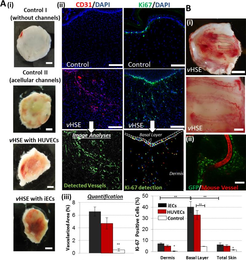
Vascularization of engineered human skin constructs is crucial for recapitulation of systemic drug delivery and for their long-term survival, functionality, and viable engraftment. In this study, the latest microfabrication techniques are used and a novel bioengineering approach is established to micropattern spatially controlled and perfusable vascular networks in 3D human skin equivalents using both primary and induced pluripotent stem cell (iPSC)-derived endothelial cells. Using 3D printing technology makes it possible to control the geometry of the micropatterned vascular networks. It is verified that vascularized human skin equivalents (vHSEs) can form a robust epidermis and establish an endothelial barrier function, which allows for the recapitulation of both topical and systemic delivery of drugs. In addition, the therapeutic potential of vHSEs for cutaneous wounds on immunodeficient mice is examined and it is demonstrated that vHSEs can both promote and guide neovascularization during wound healing. Overall, this innovative bioengineering approach can enable in vitro evaluation of topical and systemic drug delivery as well as improve the potential of engineered skin constructs to be used as a potential therapeutic option for the treatment of cutaneous wounds.

Keywords: engineered skin; iPSC; microfluidics; patterning; vasculature.


Figures

Figure 1
Figure 1. Development of vascularized HSEs
A) Schematic description of the protocol to develop vHSEs. Briefly, a sacrificial layer of alginate microchannels was created in 3D-printed molds, which had the desired vasculature pattern. Then the dermal compartment that consisted of dermal fibroblasts and collagen gel was formed around the sacrificial layer, which was suspended in the transwell inserts using a ring-shaped holder. After seeding the keratinocytes over the dermal compartment, the construct underwent epidermilization and cornification accompanied with a significant contraction. The sacrificial layer was dissolved by sodium citrate treatment through the inlet/outlet ports followed by EC seeding through the same ports. The vHSEs were then used either for grafting or in vitro perfusion experiments. B) Two different vasculature patterns were used in our studies and generated using fluorescently-tagged alginate. Scale bar: 600 μm.
Figure 2
Figure 2. Epidermal integrity and endothelial barrier function in vHSEs
A) (i) H&E and (ii) immunofluorescent staining of histological sections of vHSE generated using iPSC-derived ECs. The sections were immunolabeled with K10, K14 and loricirin (red) and CD31 (green) to evaluate epidermal integrity and endothelial coating in the microchannels. Scale bars: 250 μm. B) Evaluation of the endothelial barrier function in vHSEs. (i) The microchannels seeded with fibroblasts or iPSC-derived ECs were perfused with a fluorescently tagged dextran (70 kDa) solution. Diffusion of the dye into the collagen gel was imaged for 60 mins and fluorescence intensity was quantified at each pixel along the horizontal centerline of images using ImageJ software. (ii) Distribution of the dextran in vHSEs after 60 mins was estimated using COMSOL Multiphysics software and represented in color surface plots for the microchannels with fibroblasts and iECs. The color spectrum depicts the dextran concentration ranging from dark red (highest) to dark blue (lowest). C) Quantification of the fluorescence intensity in the microchannels and estimated values of permeability and diffusivity of the microchannels with fibroblasts, HUVECs, iECs or without cells. (**p<0.005 and N=3 HSEs)
Figure 3
Figure 3. Engraftment of vHSEs onto SCID mice
A) The effect of vHSEs on host neovascularization. Blood perfusion in HSEs (i) without channels (Control I), with acellular microchannels (Control II), vHSEs with HUVECs, and iECs grafted on SCID mice and harvested after 2 weeks. Scale bar: 2.5 mm. ii) Immunostaining of explanted HSEs (control) and vHSEs with CD31 (red) and Ki67 (green) to visualize the invasion of the host vessels and the presence of proliferating cells, respectively. Scale bars: 250 μm. iii) Quantification of the percentage of the total area covered by the host vasculature and the percentage of Ki-67 positive cells (*p<0.05, **p<0.005 and N=4). B) The effect of vasculature pattern on the host neovascularization. i) Pictures of newly formed host vasculature following the micropatterned human iEC-containing microchannels in vHSEs in comparison to the HSEs containing microchannels with no cells. Scale bars: 2.5 mm and 500 μm ii) Confocal image of the explanted vHSEs showing the overlapping pattern of Rhodamine-dextran (70kDa) perfused mice vessels and the microchannels containing GFP-tagged HUVECs. (N=4 for all conditions) Scale bar: 500 μm.

References

    1. Supp DM, Wilson-Landy K, Boyce ST. FASEB journal : official publication of the Federation of American Societies for Experimental Biology. 2002;16:797. - PMC - PubMed
    1. Maione AG, Brudno Y, Stojadinovic O, Park LK, Smith A, Tellechea A, Leal EC, Kearney CJ, Veves A, Tomic-Canic M, Mooney DJ, Garlick JA. Tissue engineering. Part C, Methods. 2015;21:499. - PMC - PubMed
    1. Bottcher-Haberzeth S, Biedermann T, Reichmann E. Burns : journal of the International Society for Burn Injuries. 2010;36:450. - PubMed
    1. Itoh M, Kiuru M, Cairo MS, Christiano AM. Proceedings of the National Academy of Sciences of the United States of America. 2011;108:8797. - PMC - PubMed
    1. Marino D, Luginbuhl J, Scola S, Meuli M, Reichmann E. Science translational medicine. 2014;6:14. - PubMed

Publication types